
Cancer Conditions
Featured Cancer Conditions
All Cancer Conditions
A Brief History of Cancer
Cancer Conditions
This history follows the research breakthroughs that built our understanding of cancer — and how steady progress in science has lowered cancer risk, found cancer sooner, improved treatment and helped people with cancer live longer. Cancer has affected animals for millions of years; scientists have found it in dinosaur bones 70 to 80 million years old. The earliest signs in humans are tumors in fossilized bones and mummies from ancient Egypt, around 3000 BCE. The oldest written description, in the Edwin Smith Papyrus from about 3000 BCE, describes a "bulging tumor of the breast." The Greek doctor Hippocrates (460–370 BCE) first used the word for cancer, calling tumors carcinos and carcinoma — Greek for crab — because they looked like a moving crab. The Roman doctor Celsus (25 BCE–50 CE) later translated this into the Latin word cancer. The Greek doctor Galen (130–200 CE) first used oncos, Greek for swelling, which gives us "oncology" and "oncologist." Ancient Egyptians blamed cancer on the gods. Hippocrates believed it came from an imbalance of body fluids. From the 1500s to the 1700s, scientists thought it might come from lymph fluid problems, long-term inflammation, injury, or an infection passed between people. Pathology and the microscope. The Italian doctor Giovanni Morgagni did the first autopsies in 1761, linking illness to cause of death; his reports began scientific oncology. The modern microscope of the 1800s showed that cancer is made of cells, that every cell comes from another cell, and that cancer cells look different from normal cells. Pathologists could then diagnose cancer in tissue removed at surgery and tell surgeons whether all of it was out. Epidemiology. In 1713, Bernardino Ramazzini noted that nuns rarely had cervical cancer but often had breast cancer, pointing toward the roles of hormones and sexually transmitted infections. In 1775, Percival Pott showed that soot caused scrotal cancer in chimney sweeps, opening the study of job-related exposures. The National Cancer Act of 1971 created a network of cancer registries and the SEER (Surveillance, Epidemiology, and End Results) Program to track cancer rates and survival. Carcinogens. Tobacco was linked to cancer in 1761. In 1911 a virus was found that caused cancer in chickens, which led to links between viruses and human cancers — hepatitis B with liver cancer, and Epstein-Barr virus with non-Hodgkin lymphoma. In the 1980s the bacterium H. pylori was shown to raise stomach cancer risk. In 1991, research showed that environmental exposures damage DNA and make cells more sensitive to carcinogens such as UV radiation and cigarette smoke. In 1999, HPV was found in 99.7% of cervical cancers, which led to the HPV vaccine in 2006. Genes. The discovery of DNA's structure in the mid-20th century showed how gene changes, inherited or caused by carcinogens, lead to cancer, and that cancer cells with damaged DNA do not die as normal cells do. In the 1970s scientists identified oncogenes and tumor suppressor genes; inherited BRCA1 and BRCA2 changes, for example, raise breast and ovarian cancer risk. The Human Genome Project, completed in 2000, helped find more cancer-related gene changes and develop better treatments. Screening tests find cancer in people without symptoms, often before it spreads, and some — for cervical and colorectal cancer — find growths before they become cancer. The Pap test, developed in the 1920s, was the first widely used screening test and cut cervical cancer rates by more than half. The discovery that HPV causes most cervical cancers led to guidelines for a primary HPV test, with or without a Pap test. The mammogram, developed in the 1960s, is still the most reliable breast cancer screening test. Colonoscopy and stool-based and blood-based tests now screen for colorectal cancer, and low-dose chest CT screens people at higher risk of lung cancer. Multi-cancer early detection (MCED) tests look for signs of many cancers in blood and other fluids; some are available, but they are expensive and not yet FDA-approved. Surgery and anesthesia. Early surgery often did more harm than good, and Galen believed cancer could not be cured. Effective anesthesia in 1846 let surgeons remove tumors and lymph nodes more completely and safely. The radical mastectomy came in the late 1800s; a century later, breast-conserving surgery proved to work as well. Today CT, MRI and PET scans locate tumors without surgery, and robotic systems operate through openings little bigger than a keyhole. Hormone therapy. In 1895, removing the ovaries was shown to slow breast cancer. In 1977 the FDA approved tamoxifen, which blocks hormone activity without surgery. For prostate cancer, surgical castration in the 1940s gave way to medicines that suppress or block male hormones. Radiation therapy. X-rays were discovered in 1895 and soon used to treat cancer. Brachytherapy (internal radiation) treated cervical and prostate cancers in the early 1900s while external beam radiation improved. Proton beams in the 1960s added precision, and conformal and intensity-modulated radiation in the 1990s spared more healthy tissue. Chemotherapy. Military research found that nitrogen mustard shrank lymphomas; it was approved in 1949. Aminopterin and methotrexate then treated childhood leukemia, and by the 1960s and 1970s chemotherapy was curing Hodgkin disease, childhood acute lymphoblastic leukemia (ALL) and testicular cancer. Stem cell and bone marrow transplants. First done in 1957, they return healthy blood stem cells after disease, chemotherapy or radiation destroys the marrow, and are now a key treatment for blood cancers. Targeted therapy, immunotherapy and precision medicine. Targeted therapies began in the 1980s; trastuzumab, approved in 1998, blocks the HER2 protein in some breast cancers. Two decades later, pembrolizumab became the first treatment approved for a tumor's genetic changes rather than where it started. Immunotherapy began with interferons for hairy cell leukemia in 1986, followed by BCG for some bladder cancers, interleukins for kidney cancer, monoclonal antibodies such as rituximab for lymphoma, cancer vaccines for prostate cancer, checkpoint inhibitors for melanoma, and cell therapies such as CAR T-cell therapy for some blood cancers and TIL therapy for some solid tumors. Precision medicine now ties these together by testing the genes and proteins in a tumor or blood to choose the treatments most likely to work. More than 18 million cancer survivors were living in the US in 2022, and the number is expected to pass 22 million by 2035. The National Coalition for Cancer Survivorship (now Cancer Nation) was founded in 1986. The National Cancer Institute created its Office of Cancer Survivorship in 1996. In 2006 the Institute of Medicine's report "From Cancer Patient to Cancer Survivor: Lost in Transition" called for transition plans and better coordination between cancer teams and primary care after treatment ends. Research continues to find new ways to prevent, find and treat cancer, to meet survivors' needs, and to improve outcomes and access to care for all groups. Current examples include how physical activity affects cancer risk, new ways to calculate breast cancer risk to personalize screening, how inadequate health insurance affects survival, and new gene tests that may personalize ovarian cancer care. Which cancer screening tests are recommended for someone of my age and risk? Is my cancer one that can be found early with screening, and how often should I be screened? Which of today's treatments — surgery, radiation, chemotherapy, hormone therapy, targeted therapy or immunotherapy — fit my cancer? Should my tumor be tested so that a precision medicine treatment can be chosen? Is there a clinical trial of a newer treatment that I could join? Source: https://www.cancer.org/cancer/understanding-cancer/history-of-cancer.html
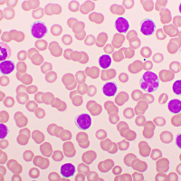
Acute Lymphocytic Leukemia (ALL) in Adults
Cancer Conditions
Get an overview of acute lymphocytic leukemia (also known as acute lymphoblastic leukemia) and the latest key statistics in the US. Acute lymphocytic leukemia (ALL) is a cancer that starts in your bone marrow (the soft inner part of certain bones, where new blood cells are made). You might also hear it called acute lymphoblastic leukemia . With this type of leukemia, the cancer cells usually invade your blood fairly quickly. They can also sometimes spread to other parts of your body, including your lymph nodes, liver, spleen, central nervous system (brain and spinal cord), testicles (in males), and other organs. What Is Cancer? Cancer starts when cells in the body begin to grow out of control. Cells in nearly any part of the body can become cancer cells. Learn more here. Leukemias are cancers that start in cells that would normally develop into different types of blood cells. Most often, leukemia starts in early forms of white blood cells, but some leukemias start in other blood cell types. There are several types of leukemia. They are divided based mainly on whether the leukemia is acute (fast growing) or chronic (slower growing), and whether it starts in myeloid cells or lymphoid cells. ALL is an acute (fast-growing) leukemia that starts in lymphoid cells. ALL is an acute leukemia; it can progress quickly and usually needs to be treated right away. It is more common in children and teens than in adults. It starts in early (immature) forms of lymphocytes , a type of white blood cell. There are 2 main types of ALL: B-cell ALL starts in early forms of B lymphocytes (B cells). T-cell ALL starts in early forms of T lymphocytes (T cells). ALL can start in early B cells or T cells at different stages of maturity. This is discussed in Acute Lymphocytic Leukemia (ALL) Subtypes and Prognostic Factors . ALL isn’t the only cancer that starts in the lymphocytes. Other types of cancer, such as lymphomas , also start in these cells. (This is true of non-Hodgkin lymphoma and Hodgkin lymphoma .) The main difference between lymphocytic (lymphoblastic) leukemias like ALL and lymphomas: Leukemias mainly affect the bone marrow and blood. Lymphomas mainly affect the lymph nodes or other organs, although they may also involve the bone marrow. But there is often some overlap. Sometimes it can be hard to tell if a cancer of lymphocytes is a leukemia or a lymphoma. In fact, the World Health Organization considers ALL and lymphoblastic lymphoma (LBL) to be different versions of the same disease (ALL/LBL). Usually, if at least 20% of the bone marrow is made up of early forms lymphocytes (called lymphoblasts or just blasts ), the disease is called ALL. If the disease is in other parts of the body with fewer blasts in the bone marrow, it is labeled as LBL. Anatomy Gallery —Blood Cells and Bone Marrow (includes more about lymphocytes) How Do Blood Cells Mature and What Happens When They Don’t Form Normally It's important to have open and honest discussions with your cancer care team about your acute lymphocytic leukemia (ALL) . Ask about anything you don’t understand or want to know more about. Below are questions to consider asking about your ALL. Can you explain what ALL is? How is it different from other types of leukemia? What subtype of ALL do I have? What does this mean? Are there other factors that might affect my prognosis ? Do I need any other tests before we can decide on treatment? Do I need to see any other types of doctors? How much experience do you and this medical center have in treating ALL? What are my treatment choices ? What do you recommend, and why? Should we consider a stem cell transplant ? When? Should I get a second opinion before starting treatment? Can you suggest a doctor or medical center? How soon do I need to start treatment? What should I do to be ready? How long will treatment last? What will it be like? Where will it be done? What are the risks and side effects of the treatments that you recommend? Could treatment affect my ability to have children ? If so, how? How will treatment affect my daily activities? What is my prognosis (outlook)? Once treatment begins, you’ll need to know what to expect and what to look for. Not all of these questions may apply to you, but getting answers to the ones that do can be helpful. How will we know if the treatment is working? What type of follow-up will I need after treatment? Is there anything I can do to manage side effects? What symptoms or side effects should I tell you about right away? How can I reach you on nights, holidays, or weekends? Do I need to change what I eat during treatment? Are there any limits on what I can do? Should I exercise? What should I do, and how often? What are my options if treatment isn’t working? Where can I find more information and support? Can you suggest a mental health professional I can see if I start to feel overwhelmed, depressed, or distressed? Be sure to write down any questions you have that aren’t on this list. For example, you might want to ask about recovery times so you can plan your work or activity schedule. Or you might want to ask if you qualify for any clinical trials . It can be hard to remember everything you’re told at each doctor’s visit, especially if you’re anxious or afraid. Even if the doctor carefully explains things, you might not hear or remember all that’s said or shown to you. Here are some ways to help you remember everything your doctor tells you. Take notes on what your doctor says. Ask if you can record your talks. Take a family member or friend with you. Ask them to take notes and remind you of questions you want to ask, so you can focus on listening and talking directly with the doctor. Doctors aren’t the only ones who can give you information. Other health care professionals, such as nurses and social workers, can answer a lot of your questions. To learn more about speaking with your health care team, see The Doctor-Patient Relationship . What Is Acute Lymphocytic Leukemia (ALL)? If you've been diagnosed with acute lymphocytic leukemia (ALL) or are worried about it, you likely have a lot of questions. Learning some basics is a good place to start. Key Statistics See the latest estimates for new cases of acute lymphocytic leukemia (ALL) and deaths in the US. What’s New in ALL Research? Research into causes, prevention, and treatment of acute lymphocytic leukemia is ongoing in many medical centers throughout the world. Find out what's new. About Acute Lymphocytic Leukemia (ALL) What’s New in Acute Lymphocytic Leukemia (ALL) Research? Key Statistics for Acute Lymphocytic Leukemia (ALL) What Is Acute Lymphocytic Leukemia (ALL)? Causes, Risk Factors, and Prevention Early Detection, Diagnosis, and Types Treating Acute Lymphocytic Leukemia (ALL) After Treatment Can Acute Lymphocytic Leukemia (ALL) Be Found Early? Signs and Symptoms of Acute Lymphocytic Leukemia (ALL) Tests for Acute Lymphocytic Leukemia (ALL) Acute Lymphocytic Leukemia (ALL) Subtypes and Prognostic Factors Chemotherapy for Acute Lymphocytic Leukemia (ALL) Targeted Therapy Drugs for Acute Lymphocytic Leukemia (ALL) Immunotherapy for Acute Lymphocytic Leukemia (ALL) Surgery for Acute Lymphocytic Leukemia (ALL) Radiation Therapy for Acute Lymphocytic Leukemia (ALL) Stem Cell Transplant for Acute Lymphocytic Leukemia (ALL) Health Professionals Who Are Part of a Cancer Care Team Source: https://www.cancer.org/cancer/types/acute-lymphocytic-leukemia.html
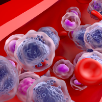
Acute Myeloid Leukemia (AML) in Adults
Cancer Conditions
Cancer starts when cells in the body begin to grow out of control. There are many kinds of cancer. Cells in nearly any part of the body can become cancer. Leukemias are blood cancers. They start in cells that would normally develop into different types of blood cells. Most often, leukemia starts in early forms of white blood cells, but some leukemias start in other blood cell types. What Is Cancer? Cancer starts when cells in the body begin to grow out of control. Cells in nearly any part of the body can become cancer cells. Learn more here. There are several types of leukemia. They are divided based mainly on whether the leukemia is acute (fast growing) or chronic (slower growing), and whether it starts in myeloid cells or lymphoid cells. Acute myeloid leukemia (AML) starts in the bone marrow, the soft inner part of certain bones, where new blood cells are made. Most often AML quickly moves from the bone marrow into the blood. It can sometimes spread to other parts of the body including the lymph nodes, liver, spleen, central nervous system (brain and spinal cord), and testicles. Sometimes leukemia cells form a tumor called a myeloid sarcoma . Different types of AML are discussed in Acute Myeloid Leukemia (AML) Subtypes and Prognostic Factors . Acute myeloid leukemia (AML) has many other names, including acute myelocytic leukemia, acute myelogenous leukemia, acute granulocytic leukemia, and acute non-lymphocytic leukemia. Most often, AML develops in early forms of myeloid cells (see below) that would normally turn into white blood cells. But it can also develop in early forms of other myeloid cells. AML tends to progress quickly, and it usually needs to be treated right away. AML is one of the most common leukemias in adults. There are different subtypes of AML. Most subtypes are classified on how mature (developed) the leukemia cells are, and how different they are from normal cells. To understand leukemia, it helps to know about the blood and lymph systems. Bone marrow is the soft inner part of certain bones. It is made up of blood-forming cells, fat cells, and supporting tissues. A small fraction of the blood-forming cells are blood stem cells . Inside the bone marrow, blood stem cells develop into new blood cells. During this process, the cells become either lymphocytes (a kind of white blood cell) or other blood-forming cells, which are types of myeloid cells . Myeloid cells can develop into red blood cells, white blood cells (other than lymphocytes), or into the cells that make platelets. Some of these myeloid cells are the ones that are abnormal in AML. There are 3 main types of blood cells: Red blood cells (RBCs) carry oxygen from the lungs to all other tissues in the body and take carbon dioxide back to the lungs to be removed. Platelets are small pieces of cells that split off from a type of bone marrow cell called a megakaryocyte . Platelets are important in stopping bleeding. They help plug up holes in blood vessels caused by cuts or bruises. White blood cells (WBCs) help the body fight infections. There are different types of WBCs, which work in slightly different ways. It's important to have open and honest communications with your cancer care team. Ask about anything you don’t understand about acute myeloid leukemia (AML) or want to know more about. For instance, consider these questions: Can you explain to me what AML is? How is it different from other types of leukemia? What subtype of AML do I have? Are there any other factors that might affect my prognosis ? Do I need any other tests before we can decide on treatment? Will I need to see any other types of doctors? Do you and this medical center have a lot of experience treating AML? What are my treatment choices? Should we consider a stem cell transplant ? When? Which treatment do you recommend, and why? Should I get a second opinion ? Can you suggest a doctor or cancer center? What should I do to be ready for treatment? How long will treatment last? What will it be like? Where will it be done? What are the risks and side effects of treatment? How long are they likely to last? How will treatment affect my daily activities? What is my prognosis (outlook)? Once treatment begins, you’ll need to know what to expect and what to look for. Not all of these questions may apply to you, but getting answers to the ones that do may be helpful. How will we know if the treatment is working? What type of follow-up will I need after treatment? Is there anything I can do to help manage side effects? What symptoms or side effects should I tell you about right away? How can I reach you on nights, holidays, or weekends? Will I be able to return to work? Should I go on disability? Do I need to eat a special diet during treatment? Are there any limits on what I can do? Should I exercise? What should I do, and how often? Can you suggest a mental health professional I can see if I start to feel overwhelmed, depressed, or distressed? What would my options be if the treatment isn’t working? Where can I find more information and support? Be sure to write down any questions you have that are not on this list. For instance, you might want specific information about expected recovery times or returning to work. Or you might want to ask if you qualify for any clinical trials . It’s hard to remember all of the things you’re told at each doctor’s visit, especially when you’re anxious or afraid. Even if the doctor carefully explains things, you might not hear or remember all that’s said or shown to you. Here are some ways to help you remember everything your doctor tells you. Take notes on what your doctor says. Ask if you can record your talks. Take a family member or friend with you. Ask them to remind you of questions you want to ask and ask them to take notes, so you can focus on listening and talking directly with the doctor. Keep in mind that doctors aren’t the only ones who can give you information. Other health care professionals, such as nurses and social workers, might be able to answer some of your questions. You can find out more about speaking with your health care team in The Doctor-Patient Relationship . About Acute Myeloid Leukemia (AML) What Is Acute Myeloid Leukemia (AML)? Key Statistics for Acute Myeloid Leukemia (AML) What’s New in Acute Myeloid Leukemia (AML) Research? Causes, Risk Factors, and Prevention Early Detection, Diagnosis, and Types Treating Acute Myeloid Leukemia (AML) After Treatment Can Acute Myeloid Leukemia (AML) Be Found Early? Signs and Symptoms of Acute Myeloid Leukemia (AML) Tests for Acute Myeloid Leukemia (AML) Acute Myeloid Leukemia (AML) Subtypes and Prognostic Factors Chemotherapy for Acute Myeloid Leukemia (AML) Targeted Therapy Drugs for Acute Myeloid Leukemia (AML) Non-Chemo Drugs for Acute Promyelocytic Leukemia (APL) Surgery for Acute Myeloid Leukemia (AML) Radiation Therapy for Acute Myeloid Leukemia (AML) Stem Cell Transplant for Acute Myeloid Leukemia (AML) Treatment of Children With Acute Myeloid Leukemia (AML) . Typical Treatment of Acute Myeloid Leukemia (Except APL) Treatment of Acute Promyelocytic Leukemia (APL) Treatment Response Rates for Acute Myeloid Leukemia (AML) If Acute Myeloid Leukemia (AML) Doesn’t Respond or Comes Back After Treatment Health Professionals Who Are Part of a Cancer Care Team Source: https://www.cancer.org/cancer/types/acute-myeloid-leukemia.html
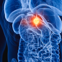
Adrenal Cancer
Cancer Conditions
The adrenals are small glands that sit on top of each of the kidneys. The kidneys are located deep inside the upper part of the abdomen. What Is Cancer? Cancer starts when cells in the body begin to grow out of control. Cells in nearly any part of the body can become cancer cells. Learn more here. Anatomy Gallery: Female Genitourinary System Explore our 3D interactive tour of the female genitourinary system. Anatomy Gallery: Male Genitourinary System Explore our 3D interactive tour of the male genitourinary system. Each adrenal gland has 2 parts. The outer part, the cortex, is where most tumors develop. The cortex makes certain hormones for the body. These hormones all have a similar chemical structure and are called steroids : Cortisol causes changes in metabolism to help the body to handle stress. Aldosterone helps the kidneys regulate the amount of salt in the blood and helps regulate blood pressure. Adrenal androgens , also called dehydroepiandrosterone or DHEA, can be converted to the sex hormones, estrogen and testosterone in other parts of the body. The amount of these hormones, however, that results from conversion of adrenal androgens is small compared to what is made in the other places in the body. In men, the testicles produce most of the androgens (male hormones). The ovaries produce most of the estrogens (female hormones) in women. The inner part of the adrenal gland, the medulla, is really an extension of the nervous system. Nervous system hormones such as norepinephrine, epinephrine, and dopamine are made in the medulla. Tumors and cancers that start in the adrenal medulla include pheochromocytomas (which most often are benign) and neuroblastomas . Tumors and cancers of the adrenal cortex are covered here, but tumors of the adrenal medulla are not. Neuroblastomas are covered separately elsewhere . Many adrenocortical tumors are discovered by accident (incidentally) when CT or MRI scans of the abdomen are done because of an unrelated health problem. These tumors are called adrenal incidentalomas , if the mass is larger than 1 cm (less than ½ inch) and is found incidentally. Regardless of how they are found, all adrenocortical tumors should be determined to be either: Adrenocortical adenomas (Benign or non-cancerous tumors that don’t spread beyond the adrenal gland.) Adrenocortical carcinomas (Malignant or cancerous tumors that can spread beyond the adrenal gland.) In general, the findings on imaging scans can help doctors understand if the tumor is an adenoma or carcinoma, based on the tumor’s size and how it looks (smooth vs. irregular, the lack or presence of fat, etc.). Most adrenal cortex tumors are benign adenomas. These tumors are usually small (less than 4 cm) and occur in only one adrenal gland. Most people with adrenal adenomas have no symptoms and don't know that they have an adrenal tumor. Adrenocortical adenomas are categorized by whether they make hormones (functional) or not (non-functional). Most adenomas are non-functional. However, the small percentage of adrenocortical adenomas that do make hormones can cause certain medical conditions, such as: Cushing’s syndrome (high levels of cortisol) Primary aldosteronism (high levels of aldosterone) Androgen and estrogen-secreting tumors The excess hormones can cause the same symptoms as those from adrenocortical carcinomas (cancers). To learn more, see Signs and Symptoms of Adrenal Cancers . Treatment : How an adrenocortical adenoma is treated depends on: If is causing symptoms, and If there are concerning findings on imaging scans If an adenoma is causing symptoms, either by making excess hormones or by its size pressing against other structures, surgery is considered. Some patients having hormone-related symptoms who can’t have a major operation because of other major health problems, might consider a drug treatment to block the production or actions of these hormones. Surgery is considered if the imaging scan shows an adenoma has concerning features, such as: Size greater than 4 cm (almost 1 ½ inches) Relatively low percentage of fat (called lipid-poor ) Irregular borders and appearance If an adenoma is not causing any symptoms, is smaller than 4 cm, and has no concerning features on scans, it is generally just watched. Based on the way it looks when the scan is repeated 3 months to 24 months after diagnosis, if the adenoma has gotten larger or is larger than 4 cm, surgery would be considered. If the tumor is unchanged and the patient does not have any symptoms from it, it might not need to be treated at all. Tumors that form from the adrenal cortex and can spread to other parts of the body are called adrenocortical carcinomas . They are also called adrenal cancers . Some of the adrenal cancers make excess hormones (functional) and can cause the same symptoms as functional adrenocortical adenomas. To learn more, see Signs and Symptoms of Adrenal Cancers . Adrenal cancers that do not make hormones (non-functional) may start causing symptoms when they grow to a certain size and press on nearby structures, causing abdominal pain. If it has spread to other parts of the body If the tumor can safely be removed If the patient can tolerate surgery If the cancer has not spread to other parts of the body, the tumor can be removed. If the tumor can safely be removed and the patient can tolerate such a procedure, then removal of the adrenal gland is commonly advised. This procedure is called an adrenalectomy . However, if the cancer does not meet all the above criteria, then an adrenalectomy is generally not advised. Sometimes, an adrenal tumor may be removed surgically to treat symptoms such as pain or high hormone levels. If surgery is not an option for you, your doctor may discuss other options such as close observation with no intervention, localized therapy such as radiation or ablation, or enrolling in a clinical trial. Most cancers found in the adrenal gland did not start there and are not primary adrenal cancers. Instead, they started in other organs or tissues and then spread (metastasized) through the bloodstream to the adrenal glands. For example, lung cancers, melanomas, and breast cancers may spread to the adrenals. When other cancers spread to the adrenals, they are not considered adrenal cancer. They are named and treated based on the place where they started. As you deal with adrenal cancer and the process of treatment, you should be able to have frank, open discussions with your cancer care team. Ask any questions, no matter how trivial they might seem. Among the questions you might want to ask are: Has the cancer spread beyond the adrenal gland? What is the stage of the cancer, and what does that mean for me? Will I need other tests before we can decide on treatment? Do I need to see any other doctors or health professionals? Is this type of adrenal cancer hereditary? Should I consider genetic testing? If I’m concerned about the costs and insurance coverage for my diagnosis and treatment, who can help me? What are my treatment choices? Does my cancer need to be treated right away, or can it be watched closely ? How much experience do you have treating this type of cancer? Should I get a second opinion ? How do I do that? What is the goal of treatment? What side effects should I expect from my treatments? What should I do to be ready for treatment? What will treatment be like? Where will treatment be done? What if I have trouble getting to and from my treatments because of transportation problems? Is my tumor secreting excessive amounts of hormones? If so, how will we treat the hormone excess? Once treatment begins, you’ll need to know what to expect and what to look for. Not all these questions may apply to you, but asking the ones that do may be helpful. What should I do to be ready for treatment? What are the treatments like? How long will treatment last? Where will the treatments be given? What risks or side effects should I be watchful for? Are there things I can do to reduce the side effects? How might treatment affect my daily activities? Can I still work full time? Are there any limits on what I can do? How can I reach someone on the team on nights, holidays, or weekends? Do I need to change what I eat during treatment? Can I exercise during treatment? If so, what kind of exercise should I do? If I start to feel overwhelmed, depressed, or distressed, can you suggest a mental health professional I can see? What if I need social support during treatment? How long will it take to recover from treatment? When can I go back to work after treatment? Do I need to follow a special diet after treatment? Are there any limits on what I can do? What side effects or symptoms should I watch for? How often will I need to have follow-up exams and imaging tests? How will we know if the cancer has come back? What should I watch for? Along with these sample questions, be sure to write down some of your own. For instance, you might want more information about recovery times so you can plan your work or activity schedule. You might also want to ask about clinical trials for which you may qualify. Doctors aren't the only ones who can give you information. Other health care professionals, such as nurses and social workers, may have the answers you seek. You can find more information about communicating with your health care team in The Doctor-Patient Relationship . About Adrenal Cancer What Is Adrenal Cancer? Key Statistics for Adrenal Cancer What's New in Adrenal Cancer Research? Causes, Risk Factors, and Prevention Early Detection, Diagnosis, and Staging Treating Adrenal Cancer After Treatment If You Have Adrenal Cancer Surgery for Adrenal Cancer Radiation Therapy for Adrenal Cancer Chemotherapy for Adrenal Cancer Immunotherapy for Adrenal Cancer Hormone Therapy for Adrenal Cancer Treatment Choices by Stage of Adrenal Cancer Health Professionals Who Are Part of a Cancer Care Team Source: https://www.cancer.org/cancer/types/adrenal-cancer.html
Advanced and Metastatic Cancer
Cancer Conditions
"Advanced" and "metastatic" are related words with different meanings. Some people use "advanced" to mean cancer that has spread; others use it for cancer that has grown locally but has not spread far. The two can mean different things for treatment and outlook, so if you are told you have advanced or metastatic cancer, ask your doctor exactly what the words mean in your case. "Advanced cancer" often means a cancer that is very unlikely to be cured: it can be treated, but it will not go away and stay away. Some advanced cancers can be controlled for a long time, like a chronic illness, and a few may still be curable. Even when it cannot be cured, treatment can sometimes shrink the cancer, slow its growth, relieve symptoms and help you live longer. Some people learn they have cancer when it is already advanced. For others, it becomes advanced years later, when it keeps growing despite treatment or spreads. Advanced cancer can be: Locally advanced — grown beyond where it started into nearby tissue or lymph nodes, but not to distant parts of the body. Some, such as locally advanced breast and prostate cancers, may be curable. Others, such as some locally advanced brain cancers, are not, because of their size or because they are close to vital organs or blood vessels. Metastatic — spread to other, often distant, parts of the body. Not every metastatic cancer is advanced: testicular cancer, for example, can spread and still be very curable because it responds well to some treatments. As advanced cancer grows it can cause symptoms. These can almost always be managed, even when the cancer itself no longer responds to treatment. Metastatic cancer has spread from where it started (the primary site). By distance, spread is called: Local — to nearby tissue or lymph nodes, an early stage of spread Regional — farther, but still in the same region of the body Distant — tumors far from the primary site By size, a spread of only a few cells is a micrometastasis , often too small to see on imaging; a larger area is sometimes called a macrometastasis. Sometimes the new tumors are already growing when cancer is first found, and sometimes a metastasis is found before the primary tumor. Then blood tests and imaging help find where it started. However far it spreads, a cancer keeps the name of where it started: breast cancer in the lungs is still breast cancer, because it is made of breast cancer cells, and treatment is based on that. Metastatic cancer has spread. Recurrence is cancer that comes back after successful treatment — in the same place (local), nearby (regional) or far away (distant). Relapse is the return of cancer or its symptoms after a period of improvement with no signs of disease. A second cancer is a new cancer, unrelated to the first and made of different cells. It can be a late effect of the first cancer or its treatment. When cancer is found in a new place, a biopsy of that area usually shows whether it is a metastasis or a second cancer. At diagnosis, exams and tests show how far the cancer has gone — this is staging. During treatment, your team watches whether treatment is working; growth or spread may mean a change of treatment. After treatment, follow-up visits for months or years, with scans, blood tests and other tests, look for signs that the cancer has returned. Advanced or metastatic cancer does not always cause symptoms. When it does, they depend on where it has spread: shortness of breath (lungs); pain, especially in bone; headache, vision problems, dizziness or seizures (brain); broken bones (bone); yellow skin and eyes (jaundice) and a swollen belly (liver). General signs include extreme tiredness or weakness (fatigue), weight loss without trying, and a new lump or swollen lymph node. These signs do not always mean the cancer has spread, but tell your care team about any symptom. When you say my cancer is advanced or metastatic, what exactly do you mean in my case? Is my cancer considered curable, controllable over the long term, or neither? What can treatment do for me now — shrink the cancer, slow it, or relieve symptoms? If cancer is found somewhere new, how will you tell whether it is a spread or a second cancer? Which symptoms should I watch for, and how can they be managed? Source: https://www.cancer.org/cancer/understanding-cancer/what-is-cancer/advanced-and-metastatic-cancer.html
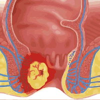
Anal Cancer
Cancer Conditions
Anal cancer is a type of cancer that starts in the anus. Cancer starts when cells in the body begin to grow out of control. To learn more about how cancers start and spread, see What Is Cancer? The anus is the opening at the lower end of the intestines. It's where the end of the intestines connect to the outside of the body. As food is digested, it passes from the stomach to the small intestine. It then moves from the small intestine into the main part of the large intestine (called the colon ). The colon absorbs water and salt from the digested food. The waste matter that's left after going through the colon is known as feces or stool . Stool is stored in the last part of the large intestine, called the rectum . From there, stool is passed out of the body through the anus as a bowel movement. The anus is connected to the rectum by the anal canal . The anal canal has two ring-shaped muscles (called sphincter muscles ) that keep the anus closed and prevent stool from leaking out. The anal canal is about 1-1/2 to 2 inches (about 3 to 5 cm) long and goes from the rectum to the anal verge . The anal verge is where the canal connects to the outside skin at the anus. This skin around the anal verge is called the perianal skin (previously called the anal margin ). The inner lining of the anal canal is the mucosa . Most anal cancers start from cells in the mucosa. Glands and ducts (tubes leading from the glands) are found under the mucosa. The glands make mucus, which acts as a lubricating fluid. Cells above the anal canal (in the rectum) and in the part of the anal canal close to the rectum are shaped like tiny columns. Most cells near the middle of the anal canal are shaped like cubes and are called transitional cells . This area is called the transitional zone . About midway down the anal canal is the dentate line , which is where most of the anal glands empty mucus into the anus. Below the dentate line are flat (squamous) cells. At the anal verge, the squamous cells of the lower anal canal merge with the skin just outside the anus. This skin around the anal verge (called the perianal skin or the anal margin ) is also made up of squamous cells, but it also contains sweat glands and hair follicles, which are not found in the lining of the lower anal canal. Anal cancers are often divided into 2 groups based on where they start: Cancers of the anal canal (above the anal verge) Cancers of the perianal skin (below the anal verge) – previously called cancers of the anal margin Sometimes, treatment may be different, depending on where the cancer is located. But anal cancers can sometimes extend from one area into the other, so it’s hard to know exactly where they started. Most (nearly 9 out of 10 cases) anal cancers in the United States are squamous cell cancer. These tumors start in the squamous cells that line most of the anal canal and the anal margin. Squamous cell cancers in the anal canal have grown beyond the surface and into the deeper layers of the lining. Squamous cell cancers of the anal margin (perianal skin) can be treated as an anal cancer or like squamous cell cancer of the skin . It is important to see an expert if you are diagnosed with perianal cancer to determine the best course of treatment. The anal cancer information here focuses mainly on anal squamous cell carcinoma since it is the most common type of anal cancer. A small number of anal cancers are adenocarcinomas . These cancers start in cells that line the upper part of the anus near the rectum. They can also start in the glands under the anal mucosa that release secretions into the anal canal. Most anal adenocarcinomas are treated the same as rectal carcinomas. For more information, see Colorectal Cancer . Adenocarcinomas can also start in apocrine glands (a type of sweat gland of the perianal skin). Paget’s disease is a type of apocrine gland carcinoma that spreads through the surface layer of the skin. Paget’s disease can affect skin anywhere in the body but most often affects skin of the perianal area, vulva , or breast . This should not be confused with Paget’s disease of the bone, which is a different disease and is not cancer. Basal cell carcinomas are a type of skin cancer that can develop in the perianal skin. These tumors are much more common in areas of skin exposed to the sun, such as the face and hands. Very few anal cancers are basal cell carcinomas. They are often treated with surgery to remove the cancer. For more information, see Skin Cancer: Basal and Squamous Cell . These cancers start in cells in the skin or anal lining that make the brown pigment called melanin . Only a very small number of anal cancers are melanomas. Melanomas are far more common on the skin in other parts of the body. If melanomas are found at an early stage (before they have grown deeply into the skin or spread to lymph nodes) they can be removed with surgery, and the outlook for long-term survival is very good. But because anal melanomas are hard to see, most are found at a later stage. If possible, the entire tumor is removed with surgery. If all of the tumor can be removed, a cure is possible. Sometimes, an abdominoperineal resection (APR) might be recommended. If the melanoma has spread too far to be removed completely, other treatments may be given. For more on this, see Melanoma Skin Cancer . These cancers are much more common in the stomach or small intestine, and rarely start in the anal region. When these tumors are found at an early stage, they are removed with surgery. If they have spread beyond the anus, they can be treated with drug therapy. For more information, see Gastrointestinal Stromal Tumor (GIST) . Some changes in the anal mucosa are harmless at first, but later might turn into cancer. These are called pre-cancers . Pre-cancers might also be called dysplasia . Some warts, for example, contain areas of dysplasia that can develop into cancer. Dysplasia in cells of the anus is called anal intraepithelial neoplasia (AIN) or anal squamous intraepithelial lesions (SILs) . Depending on how the cells look, AIN or anal SIL can be divided into 2 groups: Low-grade SIL (or grade 1 AIN): The cells in low-grade SIL look like normal cells. Low-grade SIL often goes away without treatment and has a low chance of turning into cancer. High-grade SIL (or grade 2 AIN or grade 3 AIN) : The cells in high-grade SIL look abnormal. High-grade SIL is less likely to go away without treatment and, with time, could become cancer. It needs to be watched closely and some cases of high-grade SIL need to be treated. Many types of tumors can develop in the anus. Not all of these tumors are cancers – some are benign (not cancer). Polyps are small, bumpy, or mushroom-like growths that form in the mucosa or just under it. There are many kinds including inflammatory polyps, lymphoid polyps and fibroepithelial polyps. Skin tags are benign growths of connective tissue that are covered by squamous cells. Skin tags are often mistaken for hemorrhoids (swollen veins inside the anus or rectum), but they're not the same. Anal warts (also called condylomas ) are growths that form just outside the anus and in the lower anal canal below the dentate line. Sometimes they can be found just above the dentate line. They're caused by infection with human papilloma virus (HPV). People who have or had anal warts are more likely to get anal cancer. (See “Anal conditions that are pre-cancer” above and Risk Factors for Anal cancer .) In rare cases, benign tumors can grow in other tissues of the anus. These include: Leiomyomas: Benign tumors that develop from smooth muscle cells Granular cell tumors: Tumors that develop from nerve cells and are composed of cells that contain lots of tiny spots (granules) Lipomas: Benign tumors that start from fat cells It's important to have honest, open discussions with your cancer care team. They want to answer all your questions, so that you can make informed treatment and life decisions. For instance, consider these questions: What kind of anal cancer do I have? Has my cancer spread beyond where it started? What is the stage of my cancer and what does this mean in my case? Will I need other tests before we can decide on treatment? Will I need to see other doctors? If I’m concerned about the costs and insurance coverage for my diagnosis and treatment, who can help me? How much experience do you have treating this type of cancer? Should I get a second opinion ? How do I do that? Can you recommend someone or a cancer center? What are my treatment choices? What treatment would you recommend for me? Why? What is the goal of each treatment? What are the chances my cancer can be cured with these options? How quickly do I need to decide on treatment? What should I do to be ready for treatment? How long will treatment last? What will it be like? Where will it be done? What are the risks or side effects of the treatments you suggest? How long are they likely to last? Will I need to have a colostomy? How soon do I need to start treatment? Will treatment affect my daily activities? What would my options be if the treatment doesn't work or if the cancer comes back after treatment? How will we know if the treatment is working? Is there anything I can do to help manage side effects ? What symptoms or side effects should I tell you about right away? How can I reach you on nights, holidays, or weekends? Do I need to change what I eat during treatment? Are there any limits on what I can do? Can you suggest a mental health professional I can see if I start to feel overwhelmed, depressed , or distressed ? Are there any limits on what I can do? What symptoms should I watch for? What kind of exercise should I do now? What type of follow-up will I need after treatment? How often will I need to have follow-up exams and imaging tests? Will I need any blood tests? How will we know if the cancer has come back? What should I watch for? What will my options be if the cancer comes back? How soon after treatment can I return to my normal activities, such as work, school, exercise, or sex? Along with these sample questions, be sure to write down some of your own. For instance, you might want more information about recovery times. Or you might want to ask if you qualify for a clinical trial . Doctors are not the only ones who can provide you with information. Other health care professionals, such as nurses and social workers, can also answer some of your questions. You can find out more about communicating with your health care team in The Doctor-Patient Relationship . About Anal Cancer What Is Anal Cancer? Key Statistics for Anal Cancer What’s New in Anal Cancer Research? Causes, Risk Factors, and Prevention Early Detection, Diagnosis, and Staging Treating Anal Cancer After Treatment If You Have Anal Cancer Surgery for Anal Cancer Radiation Therapy for Anal Cancer Chemotherapy for Anal Cancer Immunotherapy for Anal Cancer Treatment of Anal Cancer, by Stage Health Professionals Who Are Part of a Cancer Care Team Source: https://www.cancer.org/cancer/types/anal-cancer.html
Basal and Squamous Cell Skin Cancer
Cancer Conditions
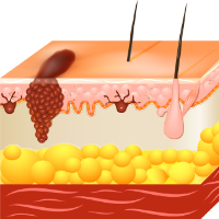
Basal and squamous cell skin cancers are the most common types of skin cancer. These cancers are often related to sun exposure. Most skin cancers start in the top layer of skin, called the epidermis . There are 3 main types of cells in this layer: Squamous cells: These are flat cells in the upper (outer) part of the epidermis, which are constantly shed as new ones form. When these cells grow out of control, they can develop into squamous cell skin cancer (also called squamous cell carcinoma ). Basal cells: These cells are in the lower part of the epidermis, called the basal cell layer. These cells constantly divide to form new cells to replace the squamous cells that wear off the skin’s surface. As these cells move up in the epidermis, they get flatter, eventually becoming squamous cells. Skin cancers that start in the basal cell layer are called basal cell skin cancers or basal cell carcinomas. Melanocytes: These cells make the brown pigment called melanin, which gives the skin its tan or brown color. Melanin acts as the body’s natural sunscreen, protecting the deeper layers of the skin from some of the harmful effects of the sun. Melanoma skin cancer starts in these cells. The epidermis is separated from the deeper layers of skin (the dermis and the subcutis) by a thin layer of tissue known as the basement membrane . When a skin cancer becomes more advanced, it generally grows through this barrier and into the deeper layers. What Is Cancer? Cancer starts when cells in the body begin to grow out of control. Cells in nearly any part of the body can become cancer cells. Learn more here. Anatomy Gallery: Skin Explore our 3D interactive tour of the skin system. Basal cell carcinoma (BCC, also called basal cell skin cancer, or just basal cell cancer ) is most common type of skin cancer. About 8 out of 10 skin cancers are basal cell carcinomas. These cancers start in the basal cell layer, which is the lower part of the epidermis. BCCs usually develop on sun-exposed areas, especially the face, head, neck, and arms. They tend to grow slowly. It’s very rare for a basal cell cancer to spread to other parts of the body. But if BCC is left untreated, it can grow into nearby areas and invade the bone or other tissues beneath the skin. If not removed completely, BCC can come back (recur) in the same place on the skin. People who have had basal cell skin cancers are also more likely to get new ones in other places. About 2 out of 10 skin cancers are squamous cell carcinomas (SCCs, also called squamous cell skin cancers, cutaneous squamous cell cancers, or just squamous cell cancers ). These cancers start in the flat cells in the upper (outer) part of the epidermis. SCCs commonly appear on sun-exposed areas of the body such as the face, ears, neck, lips, arms, and backs of the hands. They can also develop in scars or chronic skin sores elsewhere. They sometimes start in actinic keratoses (described below). Less often, they form in the skin of the genital area. Squamous cell cancers can usually be removed completely (or treated in other ways), although they are much more likely than basal cell cancers to grow into deeper layers of skin and spread to other parts of the body. Actinic keratosis (AK), also known as solar keratosis , is a pre-cancerous skin condition caused by too much exposure to the sun. AKs are usually small (less than 1/4 inch across), rough or scaly spots that may be pink-red or flesh-colored. Usually they start on the face, ears, backs of the hands, and arms of middle-aged or older people with fair skin, although they can occur on other sun-exposed areas. People who have them usually develop more than one. AKs tend to grow slowly and usually do not cause any symptoms (although some might be itchy or sore). They sometimes go away on their own, but they may come back. A small percentage of AKs may turn into squamous cell skin cancers. Most AKs do not become cancer, but it can be hard sometimes to tell them apart from true skin cancers, so doctors often recommend treating them. If they are not treated, you and your doctor should check them regularly for changes that might be signs of skin cancer. Squamous cell carcinoma in situ, also called Bowen disease, is the earliest form of squamous cell skin cancer. “In situ” means that the cells of these cancers are still only in the epidermis (the upper layer of the skin) and have not invaded deeper layers. Bowen disease appears as reddish patches. Compared with AKs, Bowen disease patches tend to be larger, redder, scalier, and sometimes crusted. Like AK, Bowen disease usually doesn’t cause symptoms, although it might be itchy or sore. Like most other skin cancers (and AKs), these patches most often appear in sun-exposed areas. Bowen disease can also occur in the skin of the anal and genital areas (where it is known as erythroplasia of Queyrat or Bowenoid papulosis ). This is often related to sexually transmitted infection with human papillomaviruses (HPVs), the viruses that can also cause genital warts. Bowen disease can sometimes progress to an invasive squamous cell skin cancer, so doctors usually recommend treating it. People who have these are also at higher risk for other skin cancers, so close follow-up with a doctor is important. Keratoacanthomas (KAs) are dome-shaped tumors that often have a crater-like area in the middle, like a volcano. These tumors tend to start on sun-exposed skin. They may start out growing quickly, but their growth usually slows down. Many keratoacanthomas shrink or even go away on their own over time without any treatment. But some continue to grow, and a few may even spread to other parts of the body. Many doctors view keratoacanthomas as a type of squamous cell skin cancer, although not all doctors agree. These tumors can be hard to tell apart from SCC just by looking at them, and their growth is often hard to predict, so doctors usually advise removing or destroying them (similar to how SCCs are treated). These cancers develop from melanocytes, the pigment-making cells in the epidermis. Melanomas are much less common than basal and squamous cell cancers, but they are more likely to grow and spread if left untreated. Melanomas are discussed in Melanoma Skin Cancer . Other types of skin cancer are much less common and are treated differently. These include: Merkel cell carcinoma Kaposi sarcoma Cutaneous (skin) lymphoma Skin adnexal tumors (tumors that start in hair follicles or skin glands) Various types of sarcomas Together, these types account for less than 1% of all skin cancers. Most skin tumors are benign (not cancerous) and rarely if ever turn into cancers. There are many kinds of benign skin tumors, including: Most types of moles (see Melanoma Skin Cancer for more about moles) Seborrheic keratoses: tan, brown, or black raised spots with a waxy texture or occasionally a slightly rough and crumbly surface when they are on the legs (also known as stucco keratosis ) Hemangiomas: benign blood vessel growths, often called strawberry spots Lipomas: soft tumors made up of fat cells Warts: rough-surfaced growths caused by some types of human papillomavirus (HPV) If you have basal or squamous cell skin cancer, it’s important to have honest, open discussions with your doctor. Ask any question, no matter how small it might seem. Here are some questions you might want to ask. What type of skin cancer do I have? Can you explain the different types of skin cancer? Has the cancer grown deeply into the skin? Has it spread to nearby areas or to other parts of the body? Do I need any other tests before we can decide on treatment? Do we need to find out the stage or risk group of the cancer? Are there any other factors that might affect my treatment options? Do I need to see any other doctors? If I’m worried about the costs and insurance coverage for my diagnosis and treatment, who can help me? How much experience do you have treating this type of cancer? What are my treatment options ? What do you recommend? Why? Will I be OK if the cancer is just removed with no other treatment? What will treatment be like? Where will it be done? What are the risks or side effects from treatment? Will I have a scar after treatment? How big will it be? How quickly do we need to decide on treatment? What should I do to be ready for treatment? What are the chances of my cancer coming back with the treatment options we have discussed? What would our options be if that happens? What are my chances of developing another skin cancer? Should I take special precautions to avoid the sun? What steps I can take to protect myself? What type of follow-up will I need after treatment? How will we know if the cancer has come back? What should I watch for? Are any of my family members at risk for skin cancer? What should I tell them to do? Along with these sample questions, be sure to write down any others you have. For instance, you might want more information about recovery times so you can plan your work or activity schedule. Or you may want to ask about second opinions or about clinical trials for which you may qualify. Keep in mind that doctors aren’t the only ones who can give you information. Other health care professionals, such as nurses and social workers, can answer some of your questions. To find out more about speaking with your health care team, see The Doctor-Patient Relationship . About Basal and Squamous Cell Skin Cancer What Are Basal and Squamous Cell Skin Cancers? Key Statistics for Basal and Squamous Cell Skin Cancers What’s New in Basal and Squamous Cell Skin Cancer Research? Causes, Risk Factors, and Prevention Early Detection, Diagnosis, and Staging Treating Basal and Squamous Cell Skin Cancer After Treatment Surgery for Basal and Squamous Cell Skin Cancers Non-surgical Local Treatments for Basal and Squamous Cell Skin Cancers Radiation Therapy for Basal and Squamous Cell Skin Cancers Systemic Chemotherapy for Basal and Squamous Cell Skin Cancers Targeted Therapy for Basal and Squamous Cell Skin Cancers Immunotherapy for Advanced Basal or Squamous Cell Skin Cancers Treating Basal Cell Carcinoma Treating Squamous Cell Carcinoma of the Skin Treating Actinic Keratosis and Bowen Disease Skin Cancer Treatments [PDF] Health Professionals Who Are Part of a Cancer Care Team Source: https://www.cancer.org/cancer/types/basal-and-squamous-cell-skin-cancer.html
Bile Duct Cancer
Cancer Conditions
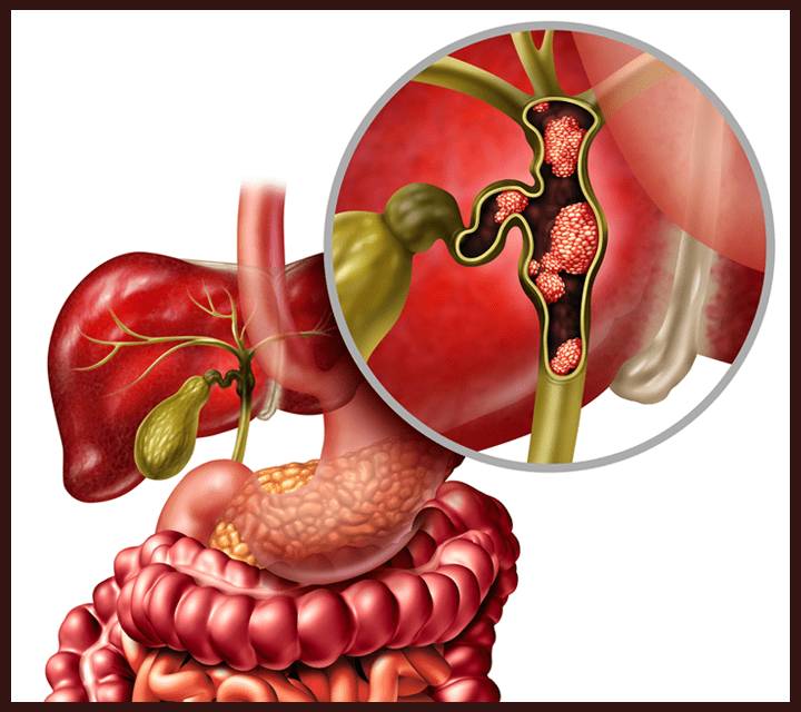
Bile duct cancer (also known as cholangiocarcinoma ) is a type of cancer that starts in a bile duct. It can start in any part of the bile duct system. To understand bile duct cancer, it helps to know about the bile ducts and what they normally do. Bile is a fluid made in your liver and stored in your gallbladder. Your bile ducts are a series of thin tubes that carry bile from your liver and gallbladder into your small intestine, where the bile helps digest the fats in food. Bile also helps your body get rid of waste material that is filtered out of the bloodstream by your liver. Different parts of the bile duct system have different names. In the liver, the bile duct system begins as many tiny tubes (called ductules ). The ductules come together to form small tubes called ducts . These small ducts merge into larger ducts and then merge again to form the left and right hepatic ducts. All of these ducts within the liver are called intrahepatic bile ducts . The left and right hepatic ducts exit the liver and join to form the common hepatic duct . Bile ducts located outside the liver are called extrahepatic bile ducts . The gallbladder is a small organ that stores bile. It is located below the liver. It is connected to the common hepatic duct via the cystic duct . These come together to form the common bile duct . The common bile duct passes through part of the pancreas before it joins with the pancreatic duct and empties into the duodenum (first part of the small intestine) at the Ampulla of Vater . What Is Cancer? Cancer starts when cells in the body begin to grow out of control. Cells in nearly any part of the body can become cancer cells. Learn more here. Anatomy Gallery: Digestive System Explore our 3D interactive tour of the digestive system. Bile duct cancer (cholangiocarcinoma) can start in any part of the bile duct system. Depending on where the cancer starts, it is either an intrahepatic cholangiocarcinoma or an extrahepatic cholangiocarcinoma . Intrahepatic bile duct cancers start in the smaller bile duct branches within the liver. They make up about 10% of all bile duct cancers. Sometimes, intrahepatic bile duct cancers are confused with hepatocellular carcinoma, a cancer that starts in the liver cells. But these are two different types of cancer, and they are treated differently. There are two types of extrahepatic cholangiocarcinoma: perihilar duct cancer and distal duct cancer. Perihilar bile duct cancers are found in the hilar region of the bile duct system. The hilar region is where the left and right hepatic ducts leave the liver and join to form the common hepatic duct. Perihilar duct cancers are also called Klatskin tumors . Distal duct cancers are found along the common bile duct. The common bile duct starts where the common hepatic duct joins with the cystic duct (from the gallbladder) and extends through the pancreas and into the small intestine at the Ampulla of Vater. Cancers that start in different parts of the bile ducts can cause different symptoms . Bile duct cancers can also be divided into types based on how the cancer cells look under the microscope. Nearly all bile duct cancers are cholangiocarcinomas (a type of adenocarcinoma, which is a cancer that starts in gland cells). Cholangiocarcinomas start in the gland cells that line the inside of the ducts. Other types of bile duct cancers are much less common. These include sarcomas, lymphomas, and small cell cancers. Our information does not cover these rare types of bile duct cancer. Not all bile duct tumors are cancer. For example, bile duct hamartomas and bile duct adenomas are types of benign (non-cancer) tumors. The most common type of cancer that starts in the liver is hepatocellular carcinoma . This type of cancer starts in the main cells that make up the liver. It is more common than cholangiocarcinoma. Cancers that start in other organs, such as the colon or rectum, can sometimes spread (metastasize) to the liver. These metastatic cancers are not true liver cancers. For example, colorectal cancer that has spread to the liver is still colorectal cancer, not liver cancer. The treatment and outlook for cancer that metastasizes to the liver is not the same as for a cancer that starts in the liver. For this reason, it’s important to know whether a tumor in the liver started in bile ducts (cholangiocarcinoma), if it started in the liver tissue itself (hepatocellular carcinoma), or if it started in another organ and has spread to the liver. It's important to have honest, open discussions with your cancer care team. They want to answer all your questions, no matter how minor they might seem. Don't be afraid to ask them. Below is a list of questions to get you started. What type of bile duct cancer do I have? Where is it located? Has my cancer spread beyond the bile ducts? What is the stage of my cancer , and what does that mean in my case? What is my prognosis (outlook)? Do I need other tests before we consider treatment options? Do I need to see any other kinds of doctors? How much experience do you have treating this type of cancer? Should I get a second opinion ? What are my treatment options ? Can my cancer be removed with surgery ? What treatment do you recommend and why? What is the goal of treatment? What risks or side effects are there to the treatments you suggest? How long are they likely to last? How quickly do we need to decide on treatment? What should I do to be ready for treatment? How long will treatment last? What will it be like? Where will it be done? How will treatment affect my daily activities? What are the chances my cancer can be cured with these treatment plans? How will we know if the treatment is working? Is there anything I can do to help manage side effects ? What symptoms or side effects should I tell you about right away? How can I reach you on nights, holidays, or weekends? Do I need to change what I eat during treatment? Are there any limits on what I can do? Can you suggest a mental health professional I can see if I start to feel overwhelmed, depressed, or distressed ? What would my options be if the treatment doesn’t work or if the cancer comes back? What type of follow-up might I need after treatment? What long-term side effects or late effects should I expect? Where can I get more information and support? Along with these sample questions, be sure to write down some of your own. For example, you might want more information about recovery times so you can plan your work or activity schedule. Or you might want to ask about qualifying for clinical trials. Keep in mind that doctors are not the only ones who can give you information. Other health care professionals , such as nurses and social workers, can answer some of your questions. Surgery for Bile Duct Cancer Radiation Therapy for Bile Duct Cancer Chemotherapy for Bile Duct Cancer Targeted Drug Therapy for Bile Duct Cancer Immunotherapy for Bile Duct Cancer Palliative Therapy for Bile Duct Cancer Treatment Options Based on the Extent of Bile Duct Cancer Health Professionals Who Are Part of a Cancer Care Team About Bile Duct Cancer Causes, Risk Factors, and Prevention Early Detection, Diagnosis, and Staging Can Bile Duct Cancer Be Found Early? Signs and Symptoms of Bile Duct Cancer Tests for Bile Duct Cancer Bile Duct Cancer Stages Questions to Ask About Bile Duct Cancer Survival Rates for Bile Duct Cancer Treating Bile Duct Cancer After Treatment If You Have Bile Duct Cancer Source: https://www.cancer.org/cancer/types/bile-duct-cancer/about/what-is-bile-duct-cancer.html
Bladder Cancer
Cancer Conditions
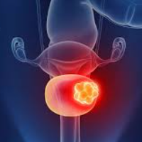
Bladder cancer starts when cells in the urinary bladder start to grow out of control. As more cancer cells develop, they can form a tumor and, with time, might spread to other parts of the body. The bladder is a hollow organ in the lower pelvis. Its main job is to store urine. Urine is liquid waste made by the kidneys and then carried to the bladder through tubes called ureters . The wall of the bladder is made up of several layers (see image). When you urinate, the muscles in the wall of the bladder contract, and urine is forced out of the bladder through a tube called the urethra . What Is Cancer? Cancer starts when cells in the body begin to grow out of control. Cells in nearly any part of the body can become cancer cells. Learn more here. Anatomy Gallery: Female Genitourinary System Explore our 3D interactive tour of the female genitourinary system. Anatomy Gallery: Male Genitourinary System Explore our 3D interactive tour of the male genitourinary system. Different types of cancer can start in the bladder. Urothelial carcinoma (transitional cell carcinoma) Urothelial carcinoma, also known as transitional cell carcinoma (TCC), starts in the urothelial cells that line the inside of the bladder. This is by far the most common type of bladder cancer. If you’re told you have bladder cancer, it's very likely to be a urothelial carcinoma. Urothelial cells also line the inside of other parts of the urinary tract, such as the part of the kidney that connects to the ureter (called the renal pelvis ), the ureters, and the urethra. People with bladder cancer sometimes have tumors in these places, too, so all of the urinary tract needs to be checked for tumors. There are different subtypes of urothelial carcinoma, which are based on how the cancer cells look under a microscope. Most often the subtype doesn’t affect how the cancer is treated, although some subtypes might be more likely to have gene changes that could affect treatment options. Sometimes urothelial cancers contain very small areas that look like some of the other cancer types below (known as divergent differentiation ). For example, the cancer may contain areas of squamous or glandular differentiation. Most often, this doesn’t affect treatment options. Other types of cancer can start in the bladder, but these are all much less common than urothelial (transitional cell) cancer. In the US, only about 3% to 5% of bladder cancers are squamous cell carcinomas (SCCs). Seen with a microscope, the cells look much like the flat cells that are found on the surface of the skin. Only about 1% to 2% of bladder cancers are adenocarcinomas. These cancers start in gland-forming cells. Less than 1% of bladder cancers are small cell carcinomas. These cancers start in nerve-like cells called neuroendocrine cells. These cancers often grow quickly and usually need to be treated with chemotherapy like that used for small cell lung cancer. Sarcomas are cancers that start in connective tissues in the body. In the bladder, sarcomas can start in the muscle cells of the bladder, although these cancers are very rare. More information on sarcomas can be found in Soft Tissue Sarcoma and Rhabdomyosarcoma. The wall of the bladder has several layers. Each layer is made up of different kinds of cells (see the image above). Most bladder cancers start in the innermost lining of the bladder, which is called the urothelium or transitional epithelium . As the cancer grows, it can invade into or through the deeper layers of the bladder wall. As the cancer becomes more advanced, it can be harder to treat. Over time, the cancer might grow outside the bladder and into nearby structures. It might spread to nearby lymph nodes, or to other parts of the body. When bladder cancer spreads, it tends to go to the lymph nodes, the bones, the lungs, or the liver. Muscle invasive vs. non-muscle invasive bladder cancer Bladder cancers are often grouped for treatment purposes based on if they have invaded into the main muscle layer of the bladder wall (see the image above): Non-muscle invasive bladder cancer (NMIBC) has not grown into the muscle layer. This is also sometimes described as superficial bladder cancer . Included in this group are both non-invasive (stage 0) bladder tumors (see below), as well as some early invasive (stage I) cancers. Muscle invasive bladder cancer (MIBC) has grown into the muscle layer of the bladder wall, and possibly deeper. These cancers are more likely to spread, and they tend to be harder to treat. In non-invasive bladder cancer, the cancer cells are still only in the inner layer (the transitional epithelium) of the bladder wall and have not grown into the deeper layers. These tumors are divided into 2 subtypes, flat and papillary, based on how they grow (see the image above). Non-invasive flat carcinomas do not grow toward the hollow part of the bladder. These tumors are also known as carcinoma in situ (CIS). Non-invasive papillary carcinomas grow in thin, finger-like projections from the inner wall of the bladder toward the hollow center. Different terms might be used to describe these tumors, based on how they look under a microscope: Papillary urothelial neoplasm of low-malignant potential (PUNLMP): These are very low-grade (slow growing) tumors. They are very unlikely to become invasive, and they tend to have very good outcomes. However, they can sometimes come back after treatment. Non-invasive low-grade papillary urothelial carcinoma (LGPUC): The cells in these tumors look a little more abnormal. While these tumors rarely become invasive, they are more likely to come back after treatment. Non-invasive high-grade papillary urothelial carcinoma (HGPUC): In these tumors, the cells look very abnormal. These tumors have a high risk of becoming invasive, and sometimes they can be a sign that there’s an invasive cancer nearby. If either a flat or papillary tumor grows into deeper layers of the bladder, it's called an invasive urothelial (or transitional cell) carcinoma. It’s important to have honest, open talks with your cancer care team. Ask any question, no matter how small it might seem. Here are some examples of things you might want to ask: What type of bladder cancer do I have? What is the stage and grade of the cancer, and what does that mean? Do you think the cancer has spread beyond my bladder? Will I need any other tests before we can decide on treatment? Do I need to see any other doctors? If I’m concerned about the costs and insurance coverage for my diagnosis and treatment, who can help me? How much experience do you have treating this type of cancer? What are my treatment options ? What do you recommend and why? What is the goal of each treatment? Should I get a second opinion ? How do I do that? Can you recommend a doctor or cancer center? What are the chances my cancer can be cured? Does my bladder need to be removed? Are there other treatment options that do not include bladder removal? If my bladder is removed, what are my options for passing urine? What are the pros and cons of each? How soon do I need to start treatment? What can I do to get ready for treatment? How long will treatment last? What will it be like? Where will it be done? What risks or side effects should I expect? How long are they likely to last? Will treatment affect my daily activities? How likely is it that the cancer will come back? Is there anything I can do to help lower this risk? What will we do if the treatment doesn’t work or if the cancer comes back? Once treatment starts, you’ll need to know what to expect and what to look for. Not all of these questions may apply, but getting answers to the ones that do may be helpful. How will we know if the treatment is working? Is there anything I can do to help manage side effects? What symptoms or side effects should I tell you about right away? How can I reach you or someone on your team on nights, holidays, or weekends? Do I need to change what I eat during treatment ? Are there any limits on what I can do? Should I exercise? What should I do, and how often? Do you know of any local or online support groups where I can talk to others who have been through this? Can you suggest a mental health professional I can see if I start to feel overwhelmed, depressed, or distressed? Are there any limits on what I can do? What kind of exercise should I do now? What type of follow-up will I need after treatment? How often will I need to have follow-up exams and tests? How will we know if the cancer has come back? What should I watch for? What would my options be if the cancer does come back? Along with these sample questions, be sure to write down any of your own. For instance, you might want to ask about recovery times so that you can plan your work or activity schedule, or you may want to ask if there are clinical trials you might be eligible for. Keep in mind that doctors aren’t the only ones who can give you information. Other health care professionals, such as nurses and social workers, can answer a lot of your questions. To find more about working with your health care team, see The Doctor-Patient Relationship . About Bladder Cancer What Is Bladder Cancer? Key Statistics for Bladder Cancer What’s New in Bladder Cancer Research? Causes, Risk Factors, and Prevention Early Detection, Diagnosis, and Staging After Treatment Treating Bladder Cancer Key Statistics for Bladder Cancer What’s New in Bladder Cancer Research? Bladder Cancer Surgery Intravesical Therapy for Bladder Cancer Chemotherapy for Bladder Cancer Radiation Therapy for Bladder Cancer Immunotherapy for Bladder Cancer Targeted Therapy Drugs for Bladder Cancer Treatment of Bladder Cancer, Based on the Stage and Other Factors Health Professionals Who Are Part of a Cancer Care Team Source: https://www.cancer.org/cancer/types/bladder-cancer.html
Blood Cancer
Cancer Conditions
Blood cancers are cancers in which different types of blood cells start to grow out of control. They can start in the bone marrow, where new blood cells are made, or in the lymphatic system, which helps the body fight infections. There are many kinds of blood cancer, including different types of leukemia and lymphoma, as well as multiple myeloma. Some grow quickly, while others develop more slowly. Some types of blood cancer affect both children and adults, especially certain types of leukemia and lymphoma. Acute Lymphocytic Leukemia (ALL) in Adults Navigate our resources on ALL (acute lymphocytic leukemia or acute lymphoblastic leukemia) from risk and prevention information to treatment guidance. Acute Myeloid Leukemia (AML) in Adults Acute myeloid leukemia is also called acute myelocytic leukemia, acute myelogenous leukemia, acute granulocytic leukemia, acute non-lymphocytic leukemia, or sometimes just AML. It is most common in older people. Childhood Leukemia Leukemia is the most common type of cancer in children and teens. Among children and teens diagnosed with cancer, 1 in 3 will have a type of leukemia. Chronic Lymphocytic Leukemia (CLL) Navigate our extensive resources on chronic lymphocytic leukemia, from risk and prevention information to treatment guidance. Chronic Myeloid Leukemia (CML) Navigate our extensive resources on CML, from risk and prevention information to treatment guidance. Chronic Myelomonocytic Leukemia (CMML) Navigate our extensive resources on CMML, from risk and prevention information to treatment guidance. Hodgkin Lymphoma Hodgkin lymphoma (also called Hodgkin disease) is a cancer of the lymph system that starts in white blood cells called lymphocytes. It often causes swollen lymph nodes, fatigue, fever, and weight loss. It is one of the most treatable cancers, especially when found early. Treatment usually includes chemotherapy, radiation, or targeted therapy. Non-Hodgkin Lymphoma Non-Hodgkin lymphoma (NHL) is a cancer that starts in white blood cells called lymphocytes, which help fight infections. It can develop in lymph nodes or other parts of the body and has many subtypes, ranging from slow-growing to aggressive forms. Symptoms may include swollen lymph nodes, fever, night sweats, and weight loss. Treatment depends on the type and stage and may include chemotherapy, targeted therapy, immunotherapy, or radiation. Non-Hodgkin Lymphoma in Children Non-Hodgkin lymphoma (NHL) is one of the most common childhood cancers (though less common than leukemia). It tends to grow quickly, but often responds well to treatment. Many children with NHL can be cured with proper treatment. Lymphoma of the Skin Lymphoma of the skin is a rare type of non-Hodgkin lymphoma that starts in the skin. It can cause red, scaly patches, lumps, or tumors on the skin. Some types grow slowly, while others spread more quickly. Treatment may include topical therapies, phototherapy, radiation, or systemic treatments like targeted therapy or chemotherapy. Waldenstrom Macroglobulinemia Waldenstrom macroglobulinemia is a rare blood cancer that affects white blood cells called B cells. These cells produce too much of a protein called macroglobulin, which can thicken the blood and cause symptoms like fatigue, bleeding, vision problems, and nerve damage. Treatment may include targeted therapy, chemotherapy, or plasma exchange to reduce thickened blood. Multiple Myeloma Multiple myeloma is a cancer that starts in plasma cells, a type of white blood cell that helps fight infections. Abnormal plasma cells build up in the bone marrow, crowding out healthy cells and causing weak bones, kidney problems, and a weakened immune system. Myelodysplastic Syndromes Myelodysplastic syndromes (MDS) are a group of blood disorders where the bone marrow doesn’t make enough healthy blood cells. This can lead to anemia, infections, and bleeding problems. MDS can sometimes turn into leukemia. Treatment may include medications, blood transfusions, or a bone marrow transplant. Quick Guide: After a Leukemia Diagnosis What to expect before, during, and after treatment for leukemia. Quick Guide: After a Lymphoma Diagnosis What to expect before, during, and after treatment for lymphoma. Anatomy Gallery: Lymphatic System Explore our 3D interactive tour of the lymphatic system. Cancer Care Toolkit Organize your cancer journey with the American Cancer Society Cancer Care Toolkit. Caregiver Resource Guide A helpful tool for people who are caring for someone with cancer. View the complete guide here. Blood Cancer Leukemia Acute Lymphocytic Leukemia (ALL) in Adults Acute Myeloid Leukemia (AML) in Adults Chronic Lymphocytic Leukemia (CLL) Chronic Myeloid Leukemia (CML) Chronic Myelomonocytic Leukemia (CMML) Childhood Leukemia Lymphoma Non-Hodgkin Lymphoma Non-Hodgkin Lymphoma in Children Hodgkin Lymphoma Multiple Myeloma Myelodysplastic Syndromes Waldenstrom Macroglobulinemia Source: https://www.cancer.org/cancer/types/blood-cancer.html
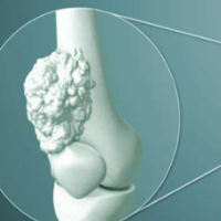
Bone Cancer
Cancer Conditions
Bone cancer is an uncommon type of cancer that begins when cells in the bone start to grow out of control. The information here focuses on primary bone cancers (cancers that start in bones) that most often are seen in adults. Information on Osteosarcoma , Ewing Tumors (Ewing sarcomas) , and Bone Metastases is covered separately. To understand bone cancer, it helps to know a little about normal bone tissue. What Is Cancer? Cancer starts when cells in the body begin to grow out of control. Cells in nearly any part of the body can become cancer cells. Learn more here. Anatomy Gallery: Skeletal System Explore our 3D interactive tour of the skeletal system. Bone is the supporting framework for your body. The hard, outer layer of bones is made of compact (cortical) bone, which covers the lighter spongy (trabecular) bone inside. The outside of the bone is covered with fibrous tissue called periosteum . Some bones have a space inside called the medullary cavity, which contains the soft, spongy tissue called bone marrow (discussed below). The tissue lining the medullary cavity is called endosteum . Most bones start out as a softer, more flexible form of tissue called cartilage . Cells in the body then lay calcium down onto the cartilage to form bone. After the bone is formed, cartilage may remain at the ends to act as a cushion between bones. This cartilage, along with ligaments and other tissues connect bones to form a joint . In adults, cartilage is mainly found at the end of some bones that are part of a joint. Cartilage can also be found in other parts of the body, such as on the ends of the ribs, in parts of the nose and ears, and in the trachea (windpipe) and larynx (voice box). Bone itself contains 2 kinds of cells. Osteoblasts are cells that lay down new bone. Osteoclasts are cells that dissolve old bone. Bone often looks as if it doesn’t change much, but it's actually very active. New bone is always forming while old bone is dissolving. This helps keep the bones strong. In some bones the marrow is only fatty tissue. In other bones it contains blood-forming cells. These cells make new red blood cells, white blood cells, and blood platelets. There are other cells in the bone marrow, too, such as plasma cells and fibroblasts . Any of these bone cells can develop into cancer. Cancers that start in the bones are called primary bone cancers . These types of cancers aren’t very common. Most of the time when an adult with cancer is told they have cancer in the bones, the doctor is talking about a cancer that started somewhere else and then spread to the bones. This is called bone metastasis. It can happen with many different types of advanced cancer , like breast cancer, prostate cancer, and lung cancer. When the cancer cells in the bone are looked at under a microscope, they look like the cancer cells in the organ they came from. So, if someone has lung cancer that has spread to bone, the cancer cells in the bone look and act like lung cancer cells, not bone cancer cells, so they need treatments that are used for lung cancer. To learn more about cancer that has spread to the bones, see Bone Metastasis . Primary bone cancers (cancers that start in the bone itself) are also known as bone sarcomas . (Sarcomas are cancers that start in bone, muscle, fibrous tissue, blood vessels, fat tissue, as well as some other tissues. They can develop anywhere in the body.) There are many kinds of primary bone cancer. Some are rare. Osteosarcoma (also called osteogenic sarcoma ) is the most common primary bone cancer. It starts in an early form of bone cells. It most often occurs in young people between the ages of 10 and 30, but about 1 in 10 osteosarcomas develop in people older than 60. It's rare in middle-aged people, and is more common in males than females. These tumors develop most often in bones of the arms, legs, or pelvis. This type of cancer is not discussed further on our Bone Cancer pages. For more information on it, see Osteosarcoma . Ewing tumors are the second most common type of primary bone cancer in children, teens, and young adults, and the third most common type of bone cancer overall. These tumors are rare in adults older than 30. They occur most often in white people and are rare among African Americans and Asian Americans. Most Ewing tumors develop in bones, but they can start in other tissues and organs. The most common sites for this cancer are the hip (pelvic) bones, the bones in the chest wall (such as the ribs or shoulder blades), the bones of the spine, and the long bones of the legs. This type of cancer is not discussed further on our Bone Cancer pages. For more information on it, see Ewing Family of Tumors . Chondrosarcoma starts in early forms of cartilage cells. It's the second most common primary bone cancer. It's rare in people younger than 20, and the risk of chondrosarcoma goes up as people get older. Chondrosarcomas can start in any place there's cartilage. Most develop in bones like the pelvic (hip) bones, legs, or arms. Some start in the trachea, larynx, chest wall, shoulder blades, ribs, or skull. Benign (non-cancerous) tumors such as enchondromas and osteochondromas are more common in the cartilage than are chondrosarcomas. These benign tumors rarely turn into cancer. People who have many of these tumors have a slightly higher chance of developing cancer, but this isn't common. Chondrosarcomas are given a grade from 1 (I) to 3 (III), which is a measure of how fast they are likely to grow. The lower the grade, the slower the cancer tends to grow and the less likely it is to spread: Low-grade (grade I) chondrosarcomas, also called atypical cartilaginous tumors , tend to grow the slowest and are very unlikely to spread. Intermediate-grade (grade II) chondrosarcomas are slightly more likely to spread. High-grade (grade III) chondrosarcomas are the most likely to spread. Most chondrosarcomas are grade I or grade II. A small number of chondrosarcomas have distinctive features that can be seen with a microscope. These uncommon subtypes often have a different prognosis (outlook): Dedifferentiated chondrosarcomas start out as typical chondrosarcomas, but then some parts of the tumor change into cells like those of a high-grade sarcoma (such as an osteosarcoma, fibrosarcoma, or high-grade undifferentiated pleomorphic sarcoma). This type of chondrosarcoma tends to develop in older adults and grows faster than most other chondrosarcomas. Mesenchymal chondrosarcomas tend to develop in younger adults. They often grow quickly, and they are more likely to come back after treatment. Clear cell chondrosarcomas are rare and tend to grow slowly. They seldom spread to other parts of the body unless they have already come back several times in the original location. This cancer was previously known as m alignant fibrous histiocytoma (MFH) of bone . Undifferentiated pleomorphic sarcoma (UPS) most often starts in soft tissues (connective tissues such as ligaments, tendons, fat, and muscle). It's rare in bones, but when it does occur, it usually affects the legs (often around the knees) or arms. This cancer most often occurs in elderly and middle-aged adults. It's rare in children. It tends to grow locally, but sometimes it can spread to distant parts of the body, like the lungs. Fibrosarcoma is another type of cancer that develops more often in soft tissues than it does in bones. It usually occurs in middle-aged adults. Bones in the legs, arms, and jaw are most often affected. This type of primary bone tumor can be either benign (not cancer) or malignant. The benign form is more common. These tumors are most common in people in their 20s and 30s. Giant cell bone tumors typically affect the legs (usually near the knees) or arms. They don’t often spread to distant parts of the body, but after surgery they can come back (even more than once) in the place where they started. With each recurrence, the tumor becomes a bit more likely to spread to other parts of the body (most often to the lungs). Rarely, a malignant giant cell bone tumor spreads to other parts of the body without first recurring locally. This uncommon type of bone tumor occurs in the bones of the spine, most often at the bottom of the spine (sacrum) or the base of the skull. It develops most often in adults older than 30. It's about twice as common in men as in women. Rarely it can develop in children, as well. Chordomas tend to grow slowly and often do not spread to other parts of the body. They often come back in the same area if they are not removed completely. If they do spread, they most often go to the lymph nodes, lungs, or liver. Some tumors that start in the bones are benign (not cancer). Benign tumors do not spread to other parts of the body and are not usually life threatening. When they need to be treated they often can be cured with surgery. Types of benign bone tumors include: Osteoid osteoma Osteoblastoma Osteochondroma Enchondroma Chondromyxoid fibroma Benign giant cell tumors of bone can also be included here. Benign bone tumors won't be discussed further here. Some other cancers develop in the bones, but they don't start in the actual bone cells. These cancers start in early forms of blood cells or immune cells in the bone marrow (the soft inner part of some bones). These are not considered to be primary bone cancers, and they are discussed in more detail elsewhere. In multiple myeloma, many tumors develop in the bones, but it's not a primary bone cancer because it starts in plasma cells (a type of immune cell) in the bone marrow. Sometimes, myeloma can be found as a single tumor (called a plasmacytoma) in a bone, but most often it has spread to other bones, so it's treated as a widespread disease. For more on this type of cancer, see Multiple Myeloma . Leukemias start in the blood-forming cells of the bone marrow, not in the bone itself. There are many types of leukemia. Most of these are cancers of early forms of white blood cells, but they can also start in other types of blood cells. For more on this type of cancer, see Leukemia . Non-Hodgkin lymphomas start in early forms of white blood cells called lymphocytes. Most often these cancers develop in lymph nodes or in other parts of the body that contain lymph tissue. But in rare cases, a lymphoma can show up first in the bones. This is known as a primary non-Hodgkin lymphoma of bone (PLB) . It can affect one or many bones. PLB is treated based on the type of lymphoma it is. (Most PLBs are diffuse large B-cell lymphomas.) The outlook is generally similar to other lymphomas of the same type and stage. For more information on the treatment of lymphoma, see Non-Hodgkin Lymphoma . It’s important to have honest, open discussions with your health care team. Ask any question, no matter how small it might seem. For instance, consider these questions: The information here focuses on primary bone cancers (cancers that start in bones) that most often are seen in adults. Information on Osteosarcoma, Ewing Tumors (Ewing sarcomas), and Bone Metastasis is covered separately. How much experience do you have doing this type of biopsy ? Are you part of a team that treats bone cancers? What will happen during the biopsy? How long will it take to get the results from the biopsy? What type of bone cancer do I have? Has the cancer spread outside the bone? What is the stage of my cancer, and what does that mean? Do you think the cancer can be resected (removed) completely? Do I need any other tests before we can decide on treatment? Will I need to see any other types of doctors? How much experience do you have treating this type of cancer? Who else will be part of the treatment team, and what do they do? What are my treatment options ? What do you recommend and why? (For tumors on an arm or leg) Which is the better surgical option: limb-sparing surgery or amputation? Why? Are there any clinical trials we should consider? How can I find out more about them? What’s the goal of treatment? Should I get a second opinion ? How do we do that? Can you recommend a doctor or cancer center? How soon do I need to start treatment? What should I do to be ready for treatment? How long will treatment last? What will it be like? Where will it be done? What risks or side effects are there to the treatments you suggest? Which side effects start shortly after treatment, and which ones might develop later on? Once treatment begins, you’ll need to know what to expect and what to look for. Not all of these questions may apply to you, but getting answers to the ones that do may be helpful. How will we know if the treatment is working? Is there anything we can do to help manage side effects? What symptoms or side effects should we tell you about right away? How can I reach you or someone on your team on nights, weekends, or holidays? Who can I talk to if I have questions about costs, insurance coverage, or social support? What are the chances of the cancer coming back with these treatment plans? What will our options be if this happens? What type of follow up and rehab will I need after treatment? Do you know of any local or online support groups where I can talk to others who have been through this? Along with these sample questions, be sure to write down some of your own. For instance, you might want more information about recovery times so that you can plan your work schedule. Keep in mind that doctors aren’t the only ones who can give you information. Other health care professionals, such as nurses and social workers, can answer some of your questions. To find more about speaking with your health care team, see The Doctor-Patient Relationship . About Bone Cancer What’s New in Bone Cancer Research? Key Statistics About Bone Cancer What Is Bone Cancer? Causes, Risk Factors, and Prevention Early Detection, Diagnosis, and Staging Treating Bone Cancer After Treatment If You Have Bone Cancer Surgery for Bone Cancer Radiation Therapy for Bone Cancer Chemotherapy for Bone Cancer Targeted Therapy and Other Drugs for Bone Cancer Treating Specific Types of Bone Cancer Health Professionals Who Are Part of a Cancer Care Team Source: https://www.cancer.org/cancer/types/bone-cancer.html
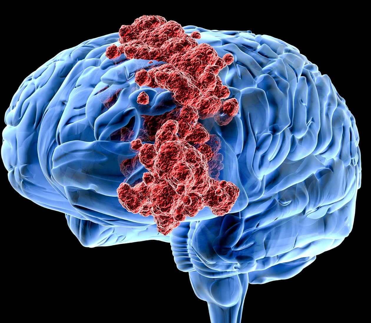
Brain and Spinal Cord Tumors in Adults
Cancer Conditions
Brain and spinal cord tumors are masses of abnormal cells in the brain or spinal cord that have grown out of control. In most other parts of the body, it is very important to distinguish between benign (non-cancerous) tumors and malignant tumors ( cancers ). Benign tumors do not grow into nearby tissues or spread to distant areas, so benign tumors in other parts of the body are almost never life-threatening. One of the main reasons malignant tumors are so dangerous is because they can spread throughout the body. Brain tumors rarely spread to other parts of the body, but most of them can spread through the brain tissue. Even so-called benign brain tumors can, as they grow, press on and destroy normal brain tissue, which can lead to serious or sometimes even life-threatening damage . For this reason, doctors usually speak of brain tumors rather than brain cancers . The main concerns with brain and spinal cord tumors are: How fast they grow How readily they spread through the rest of the brain or spinal cord Where they are located If they can be removed (or destroyed) and not come back But both benign and malignant brain and spinal cord tumors can be life threatening. Brain and spinal cord tumors tend to be different in adults and children. They often form in different areas, develop from different cell types, and may have a different outlooks and treatments. Information on these types of tumors in children is covered in Brain and Spinal Cord Tumors in Children . To understand brain and spinal cord tumors, it helps to know about the normal structure and function of the central nervous system (CNS), which is the medical name for the brain and spinal cord. The brain is the center of thought, feeling, memory, speech, vision, hearing, movement, breathing, and much more. The spinal cord and special nerves in the head called cranial nerves help carry messages between the brain and the rest of the body. These messages tell our muscles how to move, transmit information gathered by our senses, and help coordinate the functions of our internal organs. The brain is protected by the skull. Likewise, the spinal cord is protected by the bones (vertebrae) of the spinal column. The brain and spinal cord are surrounded and cushioned by a special liquid, called cerebrospinal fluid (CSF). Cerebrospinal fluid is made by the choroid plexus, which is found in spaces within the brain called ventricles . The ventricles and the spaces around the brain and spinal cord are filled with CSF. The main areas of the brain include the cerebrum, cerebellum, and brain stem. Each part has a special function. Cerebrum: The cerebrum is the large, outer part of the brain. It is divided into left and right hemispheres (halves) and controls reasoning, thought, emotion, and language. It is also responsible for planned (voluntary) muscle movements (throwing a ball, walking, chewing, etc.) and for taking in and interpreting sensory information such as vision, hearing, smell, touch, and pain. Cerebellum: The cerebellum lies under the cerebrum at the back part of the brain. It helps coordinate movement. Brain stem: The brain stem is the lower part of the brain that connects to the spinal cord. It contains bundles of very long nerve fibers that carry signals controlling muscles and sensation or feeling between the cerebrum and the rest the body. Special centers in the brain stem also help control breathing and the beating of the heart. Also, most cranial nerves (described below) start in the brain stem. The brain stem is divided into 3 main parts: the midbrain, pons, and medulla oblongata. Because the brain stem is a small area that is so essential for life, it might not be possible to surgically remove tumors in this area. Cranial nerves: The cranial nerves extend directly out of the base of the brain (as opposed to coming out of the spinal cord). These nerves carry signals directly between the brain and the face, eyes, ears, tongue, mouth, and some other areas. Spinal cord: The spinal cord is made up of bundles of very long nerve fibers that carry signals related to muscle control, sensation or feeling, and bladder and bowel control. Pituitary gland and hypothalamus: The pituitary is a small gland at the base of the brain. It is connected to a part of the brain called the hypothalamus . They work together to make hormones that help regulate the activity of several other glands in the body. For example: They control the amount of thyroid hormone made by the thyroid gland. They affect breast milk production and release. They control the amount of male or female hormones made by the testicles or ovaries. They make growth hormone, which stimulates body growth. They make vasopressin, which regulates water balance by the kidneys. The growth of tumors in or near the pituitary or hypothalamus, as well as surgery and/or radiation therapy in this area, can affect these functions. For example, tumors starting in the pituitary gland sometimes make too much of a certain hormone, which can cause problems. On the other hand, a person may have low levels of one or more hormones after treatment and may need to take hormones to make up for this. Pineal gland: The pineal gland is not really part of the brain. It is a small endocrine gland that sits between the cerebral hemispheres. It makes melatonin, a hormone that regulates sleep, in response to changes in light. The most common tumors of the pineal gland are called pineoblastomas . Blood-brain barrier: The inner lining of the small blood vessels (capillaries) in the brain and spinal cord creates a very selective barrier between the blood and the tissues of the central nervous system. This barrier normally helps maintain the brain’s metabolic balance and keeps harmful toxins from getting into the brain. Unfortunately, it also keeps out most chemotherapy drugs that are used to kill cancer cells, which in some cases limits their usefulness. Choroid plexus: The choroid plexus is the area of the brain within the ventricles that makes CSF, which nourishes and protects the brain. The brain and spinal cord have many kinds of tissues and cells, which can develop into different types of tumors . Neurons (nerve cells): These are the cells in the brain that help determine thought, memory, emotion, speech, muscle movement, sensation, and just about everything else that the brain and spinal cord do. Unlike many other types of cells that can grow and divide to repair damage from injury or disease, neurons in the brain and spinal cord largely stop dividing about a year after birth (with a few exceptions). Neurons do not usually form tumors, but they are often damaged by tumors that start nearby. Glial cells: Glial cells are the supporting cells of the brain. Most brain and spinal cord tumors develop from glial cells. These tumors are sometimes referred to as gliomas . Astrocytes help support and nourish neurons. When the brain is injured, astrocytes form scar tissue that helps repair the damage. The main tumors starting in these cells are called astrocytomas or glioblastomas . Oligodendrocytes make myelin, a fatty substance that surrounds and insulates the nerve cell axons of the brain and spinal cord. This helps neurons send electric signals through the axons. Tumors starting in these cells are called oligodendrogliomas . Ependymal cells line the ventricles (fluid-filled areas) within the brain and form part of the pathway through which CSF flows. Tumors starting in these cells are called ependymomas . (A fourth type of cell, called microglia , are the infection-fighting cells of the central nervous system. They are part of the immune system and are not truly glial cells.) Neuroectodermal cells: These are very early forms of nervous system cells that are probably involved in brain cell development. They are found throughout the brain, although they are not often seen in the adult central nervous system. The most common tumors that come from these cells develop in the cerebellum and are called medulloblastomas . Meninges: These are layers of tissue that line and protect the brain and spinal cord. CSF travels through spaces formed by the meninges. The most common tumors that start in these tissues are called meningiomas . It’s important for you to be able to have honest, open discussions with your cancer care team. Ask any question, no matter how small it might seem. Here are some you might want to ask, but be sure to add your own questions as you think of them. What kind of tumor do I have? Is the tumor benign or malignant? What does this mean? Where in the brain or spinal cord is the tumor? Has it grown into nearby areas? Will I need any other tests before we can decide on treatment? Will I need to see any other types of doctors? How much experience do you have treating this type of tumor? What are my treatment choices ? What do you recommend? Why? Should I get a second opinion ? Can you recommend a doctor or treatment center? How soon do we need to start treatment ? What’s the goal of treatment (cure, prolonging life, relieving symptoms, etc.)? How likely is it that the tumor can be removed (or destroyed) completely? Will treatment relieve any of the symptoms I now have? What are the possible risks or side effects of treatment? What disabilities might I develop? What should I do to be ready for treatment? How long will treatment take? What will it be like? Where will it be given? What is my expected prognosis (outlook)? If I'm concerned about costs and insurance coverage for my diagnosis and treatment, who can help me? Once treatment begins, you’ll need to know what to expect and what to look for. Not all of these questions might apply to you, but getting answers to the ones that do may be helpful. How will we know if the treatment is working (or has worked)? Is there anything I can do to help manage side effects ? What symptoms or side effects should I tell you about right away? How can I reach you or someone from your office on nights, holidays, or weekends? Are there any limits on what I can do? Can you suggest a mental health professional I can see if I start to feel overwhelmed, depressed, or distressed? Are there any limits on what I can do? What symptoms should I watch for? What type of follow-up will I need after treatment? How often will I need to have follow-up exams and tests? How will we know if the tumor has come back? What should I watch for? Where can I find more information and support? Along with these sample questions, be sure to write down any others you want to ask. For instance, you might want information about recovery times so that you can plan your work or activity schedule. Or you might want to ask about clinical trials that might be right for you. Keep in mind that doctors aren’t the only ones who can give you information. Other health care professionals, such as nurses and social workers, can answer some of your questions. To learn more about speaking with your health care team, see Talking With Your Doctor . About Brain and Spinal Cord Tumors in Adults What Are Adult Brain and Spinal Cord Tumors? Types of Brain and Spinal Cord Tumors in Adults Key Statistics for Brain and Spinal Cord Tumors What’s New in Adult Brain and Spinal Cord Tumor Research? Causes, Risk Factors, and Prevention Early Detection, Diagnosis, and Staging Treating Brain and Spinal Cord Tumors in Adults After Treatment If You're an Adult with a Brain or Spinal Cord Tumor Surgery for Adult Brain and Spinal Cord Tumors Radiation Therapy for Adult Brain and Spinal Cord Tumors Chemotherapy for Adult Brain and Spinal Cord Tumors Targeted Drug Therapy for Adult Brain and Spinal Cord Tumors Other Drug Treatments for Adult Brain and Spinal Cord Tumors Tumor Treating Fields (TTF) Therapy for Adult Brain and Spinal Cord Tumors Treatment of Adult Brain and Spinal Cord Tumors, by Type Health Professionals Who Are Part of a Cancer Care Team Source: https://www.cancer.org/cancer/types/brain-spinal-cord-tumors-adults.html
Brain and Spinal Cord Tumors in Children
Cancer Conditions
If your child has a brain or spinal cord tumor, knowing what to expect can help you cope. Here you can find out all about brain and spinal cord tumors in children, including risk factors, symptoms, and how they are found and treated. (For information on adult tumors see Brain and Spinal Cord Tumors in Adults .) Brain and spinal cord tumors are masses of abnormal cells in the brain or spinal cord that have grown out of control. In most other parts of the body, there's an important difference between benign (non-cancerous) tumors and malignant tumors ( cancers ). Benign tumors do not invade nearby tissues or spread to distant areas, and are almost never life threatening in other parts of the body. Malignant tumors (cancers) are so dangerous mainly because they can spread throughout the body. Brain tumors rarely spread to other parts of the body, though many of them are considered malignant because they can spread through the brain and spinal cord tissue. But even so-called benign tumors can press on and destroy normal brain tissue as they grow, which can lead to serious or sometimes even life-threatening damage. Because the difference between benign and malignant tumors isn't as important in the brain, doctors usually speak of “brain tumors” rather than “brain cancers.” How fast they grow How readily they spread through the rest of the brain or spinal cord If they can be removed or treated and not come back Both benign and malignant tumors can be life threatening. Brain and spinal cord tumors in children tend to be different from those in adults . They often form in different places, develop from different cell types, and may have a different treatment and prognosis (outlook). To learn more about the differences between childhood cancers and adult cancers in general, see Childhood Cancer. To understand brain and spinal cord tumors, it helps to know about the normal structure and function of the central nervous system (CNS), which is the medical name for the brain and spinal cord. The brain is the center of thought, feeling, memory, speech, vision, hearing, movement, and much more. The spinal cord and special nerves in the head, called cranial nerves , carry messages between the brain and the rest of the body. These messages tell our muscles how to move, transmit information gathered by our senses, and help coordinate the functions of our internal organs. The brain is protected by the skull. Likewise, the spinal cord is protected by the bones (vertebrae) of the spinal column. The brain and spinal cord are surrounded and cushioned by a liquid called cerebrospinal fluid (CSF). Cerebrospinal fluid is made by the choroid plexus, which is in spaces in the brain called ventricles . The ventricles and the spaces around the brain and spinal cord are filled with CSF. The main areas of the brain include the cerebrum, cerebellum, and brain stem. Each area has a special function. Cerebrum: The cerebrum is the large, outer part of the brain. It is made up of 2 hemispheres (halves) and controls reasoning, thought, emotion, and language. It is also responsible for planned (voluntary) muscle movements (throwing a ball, walking, chewing, etc.) and for taking in and interpreting sensory information such as vision, hearing, smell, touch, and pain. Cerebellum: The cerebellum lies under the cerebrum at the back part of the brain. It helps coordinate movement. Brain stem: The brain stem is the lower part of the brain that connects to the spinal cord. It has bundles of very long nerve fibers that carry signals controlling muscles and sensation or feeling between the cerebrum and the rest of the body. Special centers in the brain stem also help control breathing and the heart beating. In addition, most cranial nerves (described below) start in the brain stem. The brain stem is divided into 3 main parts: the midbrain, pons, and medulla oblongata. Because the brain stem is a small area that is so essential for life, it might not be possible to surgically remove tumors in this area. Cranial nerves: The cranial nerves extend directly out of the base of the brain (as opposed to coming out of the spinal cord). These nerves carry signals directly between the brain and the face, eyes, tongue, mouth, and some other areas. The most common cranial nerve tumors in children are called optic gliomas , which are tumors of the optic nerve (the large nerve that runs between the brain and each eye). Spinal cord: The spinal cord has bundles of very long nerve fibers that carry signals that control muscles, sensation or feeling, and bladder and bowel control. The brain and spinal cord have many kinds of tissues and cells, which can develop into different types of tumors . Neurons (nerve cells): These are the cells in the brain that help determine thought, memory, emotion, speech, muscle movement, sensation, and just about everything else that the brain and spinal cord do. They do this by transmitting chemical and electric signals through their nerve fibers (axons). Axons in the brain tend to be short, while those in the spinal cord can be as long as several feet. Unlike many other types of cells that can grow and divide to repair damage from injury or disease, neurons in the brain and spinal cord largely stop dividing about a year after birth (with a few exceptions). Neurons do not usually form tumors, but they can be damaged by tumors that start nearby. Glial cells: Glial cells are the supporting cells of the brain. Most brain and spinal cord tumors develop from glial cells. These tumors are sometimes referred to as a group called gliomas . Astrocytes help support and nourish neurons. When the brain is injured, astrocytes form scar tissue that helps repair the damage. The main tumors starting in these cells are called astrocytomas or glioblastomas . Oligodendrocytes make myelin, a fatty substance that surrounds and insulates the nerve cell axons of the brain and spinal cord. This helps neurons send electric signals through the axons. Tumors starting in these cells are called oligodendrogliomas. Ependymal cells line the ventricles (fluid-filled areas) within the central part of the brain and form part of the pathway through which cerebrospinal fluid (CSF) flows. Tumors starting in these cells are called ependymomas . (A fourth type of cell, called microglia, are the infection-fighting cells of the central nervous system. They are part of the immune system and are not truly glial cells.) Neuroectodermal cells: These are very early forms of nervous system cells that are probably involved in brain cell development. They are found throughout the brain. The most common tumors that come from these cells are called medulloblastomas , which start in the cerebellum. Meninges: These are layers of tissue that cover and protect the brain and spinal cord. The meninges help form the spaces through which CSF travels. The most common tumors that start in these tissues are called meningiomas . Choroid plexus: The choroid plexus is the area of the brain within the ventricles that makes CSF, which nourishes and protects the brain. Tumors that start here include choroid plexus papillomas and choroid plexus carcinomas . Pituitary gland and hypothalamus: The pituitary is a small gland at the base of the brain. It is connected to a part of the brain called the hypothalamus . Both make hormones that help regulate the activity of several other glands in the body. For example, they control the amount of thyroid hormone made by the thyroid gland, the production and release of milk by the breasts, and the amount of male or female hormones made by the testicles or ovaries. They also make growth hormone, which stimulates body growth, and vasopressin, which regulates water balance by the kidneys. The growth of tumors in or near the pituitary or hypothalamus, as well as surgery and/or radiation therapy in this area, can affect these functions. For example, tumors starting in the pituitary gland sometimes make too much of a certain hormone, which can cause problems. On the other hand, a child may have low levels of one or more hormones after treatment and may need to take hormones to make up for this. Pineal gland: The pineal gland is not really part of the brain. It is a small endocrine gland that sits between the cerebral hemispheres. It makes melatonin, a hormone that regulates sleep, in response to changes in light. The most common tumors of the pineal gland are called pineoblastomas . Blood-brain barrier: The inner lining of the small blood vessels (capillaries) in the brain and spinal cord creates a very selective barrier between the blood and the tissues of the central nervous system. This barrier normally helps maintain the brain’s metabolic balance and keeps harmful toxins from getting into the brain. Unfortunately, it also keeps out most chemotherapy drugs that are used to kill cancer cells, which in some cases limits their usefulness. Many different types of tumors can occur in the brain and spinal cord. Several factors are important when doctors are trying to figure out how best to treat a tumor and what the prognosis (outlook) is likely to be. The type of tumor (based on the type of cell it starts from): Tumors can form in almost any type of tissue or cell in the brain or spinal cord . Some tumors have a mix of cell types. Different types of tumors tend to start in certain parts of the brain or spinal cord, and tend to grow in certain ways. (The most common types of brain and spinal cord tumors in children are described below.) The grade of the tumor: Some types of brain and spinal cord tumors are more likely to grow into nearby tissues (and to grow quickly) than are others. Brain and spinal cord tumors are typically divided into 4 grades (using Roman numerals I to IV), based largely on how the tumor cells look under a microscope. The higher the grade, the more quickly the tumor is likely to grow: Lower grade (grade I or II) tumors tend to grow more slowly and are less likely to grow into (invade or infiltrate) nearby tissues. Higher grade (grade III or IV) tumors tend to grow quickly and are more likely to grow into nearby tissues. These tumors often require more intensive treatment. Gene changes in the tumor cells: Even for a specific type of tumor, the changes in the genes of the tumor cells can be different. For example, many types of tumors are now divided based on whether the cells have mutations in one of the IDH genes. For a specific type of tumor, those with IDH mutations tend to have a better outlook than those without a mutation. Other gene mutations can also be important for certain types of tumors. The location of the tumor: Where the tumor is in the brain and spinal cord can affect what symptoms it causes, as well as which treatments might be best. Brain tumors in children are more likely to start in the lower parts of the brain, such as the cerebellum and brain stem, than they are in adults. But they can start in the upper parts of the brain as well. Gliomas are not a specific type of tumor. Glioma is a general term for a group of tumors that start in glial cells (the supporting cells of the brain). A number of tumors can be considered gliomas, including: Astrocytomas (which include glioblastomas) Oligodendrogliomas Ependymomas Brain stem gliomas Optic gliomas About half of all brain and spinal cord tumors in children are gliomas. Astrocytomas are tumors that start in cells called astrocytes , a kind of glial cell that helps support and nourish nerve cells. Some astrocytomas can spread widely throughout the brain and blend with the normal brain tissue, which can make them hard to remove by surgery. Sometimes they spread along the cerebrospinal fluid (CSF) pathways. It is very rare for them to spread outside of the brain or spinal cord. Low-grade (grade I or II) astrocytomas tend to grow slowly and are the most common type in children. Some types, known as non-infiltrating astrocytomas , are grade I tumors that tend to grow very slowly and do not grow into (infiltrate) nearby tissues, so they often have a good prognosis. Pilocytic astrocytomas are grade I tumors that tend to grow slowly and rarely grow into nearby tissues. They most commonly occur in the cerebellum but can also begin in the optic nerve, hypothalamus, brain stem, or other areas. They account for nearly 1 out of 5 brain tumors in children. Subependymal giant cell astrocytomas (SEGAs) occur in the ventricles (spaces in the brain). They are grade I tumors that tend to grow slowly and rarely grow into nearby tissues. These tumors are almost always linked with an inherited condition called tuberous sclerosis . Diffuse astrocytomas are also slow-growing tumors, but they are grade II tumors that can grow into nearby tissues, which makes them hard to remove with surgery. Though these tumors are thought of as low grade, they tend to become more aggressive and fast growing over time. Pleomorphic xanthoastrocytomas (PXAs) are grade II tumors that tend to grow slowly, and most can be cured by surgery alone. Optic gliomas are astrocytomas that start in the optic nerves (the nerves leading from the eyes to the brain). They usually grow slowly, and are often linked with an inherited condition called neurofibromatosis type 1 . These tumors are rarely fatal, but they may cause vision loss and injury to nearby brain tissue. High-grade (grade III or IV) astrocytomas tend to grow quickly and spread into the surrounding normal brain tissue. These include: Glioblastomas , which are the fastest growing type of astrocytoma (grade IV). Anaplastic astrocytomas , which are grade III. These tumors start in brain cells called oligodendrocytes (a type of glial cell that makes a fatty substance that helps nerve cells send electric signals). These are grade II tumors that tend to grow slowly, but most of them can grow into nearby brain tissue and can’t be removed completely by surgery. Oligodendrogliomas rarely spread along the CSF pathways and even less frequently spread outside the brain or spinal cord. As with astrocytomas, they can become more aggressive over time. Only about 1% of brain tumors in children are oligodendrogliomas. About 5% of brain tumors in children are ependymomas. These tumors start in the ependymal cells that line the ventricles or central canal of the spinal cord. They can range from fairly low-grade (slow growing) tumors to grade III (fast growing) tumors, which are called anaplastic ependymomas . Ependymomas may spread along the CSF pathways but do not spread outside the brain or spinal cord. These tumors can block the flow of CSF out of the ventricles, causing the ventricles to become very large – a condition called hydrocephalus . Unlike astrocytomas and oligodendrogliomas, ependymomas usually do not grow into normal brain tissue. As a result, some (but not all) ependymomas can be removed and cured by surgery. But because they can spread along ependymal surfaces and CSF pathways, treating them can sometimes be difficult. A brain stem glioma is any type of glioma that starts in the brain stem. This term refers to the location of the tumor, rather than the type of cell it starts in. A small number of brain stem gliomas occur as tumors with very distinct edges (called focal brain stem gliomas ). More often, brain stem gliomas grow diffusely throughout the brain stem (where the tumor cells are spread throughout normal tissue), rather than growing as a focal tumor (where the tumor cells are clustered together). These are referred to as diffuse midline gliomas . These tumors most often start in the pons, where they are called diffuse intrinsic pontine gliomas (DIPGs) . These tumors can be hard to treat. About 10% to 20% of brain tumors in children are brain stem gliomas. Nearly all of these tumors are some type of astrocytoma. These tumors start in early forms of nerve cells in the central nervous system. About 10% to 20% of brain tumors in children are embryonal tumors. They are more common in younger children than older ones, and are rare in adults. Embryonal tumors tend to grow quickly and often spread throughout the CSF pathways. Medulloblastomas are the most common type of embryonal tumor. These tumors start in the cerebellum. There are several different types of medulloblastomas, based on how the tumor cells look under a microscope, and on which gene mutations the cells have. Some types of medulloblastoma tend to have a better outlook than others, and doctors are now trying to determine how this might affect treatment. Medulloblastomas can often be treated effectively and tend to have a better outlook than embryonal tumors in other parts of the brain. Medulloepithelioma Atypical teratoid/rhabdoid tumor (ATRT) Embryonal tumor with multilayered rosettes In the past, many embryonal tumors were referred to as primitive neuroectodermal tumors (PNETs) . Some types of tumors occur in the pineal gland (a small gland in the middle of the brain). The most common (and fastest growing) of these are called pineoblastomas . These tumors can be hard to treat. Germ cell tumors , which are described below, can also start in the pineal gland. These slow-growing tumors start above the pituitary gland but below the brain itself. They account for about 4% of brain tumors in children. These tumors may press on the pituitary gland and the hypothalamus, causing hormone problems. Because craniopharyngiomas start very close to the optic nerves, they can also cause vision problems. This makes them hard to remove completely without damaging the child’s vision or hormone balance. Certain tumors that develop in children and young adults (and rarely in older adults) have both glial and neuronal cell components. They tend to have a fairly good outlook. Dysembryoplastic neuroepithelial tumors (DNETs) tend to be slow growing (grade II) tumors, and most can be cured by surgery alone. Ganglioglioma is a type of grade I tumor that has both mature neurons and glial cells. Most can be cured by surgery alone or surgery combined with radiation therapy. These rare tumors start in the choroid plexus, the area that makes cerebrospinal fluid (CSF) within the ventricles of the brain. Most are benign ( choroid plexus papillomas ) and can be cured by surgery. However, some are malignant ( choroid plexus carcinomas ). These tumors start in Schwann cells that surround and insulate cranial nerves and other nerves. Schwannomas are usually benign . They often form near the cerebellum on the cranial nerve responsible for hearing and balance, in which case they are called vestibular schwannomas or acoustic neuromas . They may also develop on spinal nerves, just past the point where the nerve leaves the spinal cord. When this is the case, the tumor can press on the spinal cord, causing weakness, sensory loss, and bowel and bladder problems. These tumors are rare in children. When schwannomas are found in a child, particularly if there are tumors on both sides of the head, it often means the child has an inherited tumor syndrome such as neurofibromatosis type 2. (See Risk Factors for Brain and Spinal Cord Tumors in Children .) These tumors begin in the meninges, the layers of tissue that surround the outer part of the brain and spinal cord. Meningiomas cause symptoms by pressing on the brain or spinal cord. They are much less common in children than in adults. Meningiomas are almost always benign and are usually cured by surgery. Some, however, are located very close to vital structures in the brain and can’t be cured by surgery alone. Meningiomas are often assigned a grade based on how the tumor cells look. Grade I meningiomas , which look most like normal cells, account for most meningiomas. Grade II (atypical) meningiomas look slightly more abnormal. Grade III (anaplastic or malignant) meningiomas , which look the most abnormal, make up only about 1% to 3% of meningiomas. Higher-grade meningiomas are more likely to come back after treatment, and some grade III meningiomas can spread to other parts of the body. These tumors start in the bone at the base of the skull or at the lower end of the spine. Chordomas don’t start in the central nervous system, but they can injure nearby parts of the brain or spinal cord by pressing on them. These tumors tend to come back if they are not removed completely, causing more damage. They usually do not spread to other organs. Chordomas are much more common in adults than in children. For more on these tumors, see Bone Cancer . These rare tumors develop from germ cells, which normally form egg cells in women and sperm cells in men. During normal development before birth, germ cells travel to the ovaries or testicles and develop into egg or sperm cells. But sometimes some germ cells don’t move where they should and end up in abnormal locations such as the brain. They may then develop into germ cell tumors, similar to those that can form in the ovaries or testicles. Germ cell tumors of the nervous system usually occur in children, most often in the pineal gland or above the pituitary gland. These tumors can sometimes be diagnosed without a biopsy by measuring certain chemicals in the cerebrospinal fluid (CSF) or blood. Germinoma s (the most common type of CNS germ cell tumor) Choriocarcinomas Embryonal carcinomas Teratomas Yolk sac tumors (endodermal sinus tumors) These nerve cell tumors are the third most common cancer in children. But neuroblastomas rarely develop in the brain or spinal cord; most develop from nerve cells inside the abdomen or chest. This type of cancer is most common during early infancy. For more information, see Neuroblastoma . Lymphomas are cancers that start in cells called lymphocytes, which are white blood cells that are part of the immune system. Most lymphomas start in other parts of the body, but a small portion start in the central nervous system (CNS), and are called primary CNS lymphomas . These tumors are rare in children. For more on childhood lymphomas, see Non-Hodgkin Lymphoma in Children . Tumors that start in the pituitary gland are almost always benign (non-cancerous). But they can still cause problems if they grow large enough to press on nearby structures or if they make too much of any kind of hormone. These tumors are more common in teens than in younger children. For more information, see Pituitary Tumors . Sometimes tumors in the brain are found to have metastasized (spread) there from some other part of the body. Tumors that start in other organs and then spread to the brain are called metastatic or secondary brain tumors (as opposed to primary brain tumors, which start in the brain). This is important because metastatic and primary brain tumors are often treated differently. In children, metastatic brain tumors are much less common than primary brain tumors. Childhood leukemias can sometimes spread to the CSF around the brain and spinal cord. When this happens, the cancer is still considered a leukemia (the cancer cells in the CSF are leukemia cells), so doctors use treatments directed at the leukemia. For more information, see Childhood Leukemia . It’s important for you to have honest, open discussions with your child’s cancer care team. They want to answer all of your questions, no matter how minor you might think they are. Here are some questions you might want to ask. Be sure to add your own questions as you think of them. What kind of tumor does my child have? What does this mean? Where is the tumor located? Has it grown into nearby areas? Will my child need other tests before we can decide on treatment? Will we need to see any other types of doctors? How much experience do you have treating this type of tumor? What are our treatment options ? What do you recommend? Why? Should we get a second opinion ? Can you recommend a doctor or cancer center? How soon do we need to start treatment? What should we do to be ready for treatment? How long will treatment last? What will it be like? Where will it be done? How might treatment affect our daily activities? What are the possible risks and side effects of treatment? How might treatment affect my child’s ability to learn, grow, and develop? How likely is it that treatment could affect my child’s future ability to have children? Based on what you’ve learned about my child’s tumor, what is the expected prognosis (outlook)? Once treatment begins, you’ll need to know what to expect and what to look for. Not all of these questions may apply, but getting answers to the ones that do may be helpful. How will we know if the treatment is working? Is there anything we can do to help manage side effects? What symptoms or side effects should we tell you about right away? How can we reach someone from your office on nights, holidays, or weekends? Are there any limits on what my child can do? What type of follow-up will we need after treatment? How often will my child need to have follow-up exams and tests? Are there any limits on what my child can do? How will we know if the tumor has come back? What should we watch for? Are there nearby support groups or other families who have been through this that we could talk to? Along with these sample questions, be sure to write down any others you might want to ask. For instance, you might want information about recovery times so you can plan your work and your child’s school and activity schedule. Or you may want to ask about clinical trials for which your child may qualify. Also keep in mind that doctors aren't the only ones who can give you information. Other members of the treatment team, such as nurses and social workers, can answer some of your questions. You can find out more about speaking with your child’s health care team in The Doctor-Patient Relationship . About Brain and Spinal Cord Tumors in Children What Are Brain and Spinal Cord Tumors in Children? Types of Brain and Spinal Cord Tumors in Children Key Statistics for Brain and Spinal Cord Tumors in Children What’s New in Research for Childhood Brain and Spinal Cord Tumors? Causes, Risk Factors, and Prevention Early Detection, Diagnosis, and Staging Treating Brain and Spinal Cord Tumors in Children After Treatment If Your Child Has a Brain or Spinal Cord Tumor Surgery for Brain and Spinal Cord Tumors in Children Radiation Therapy for Brain and Spinal Cord Tumors in Children Chemotherapy for Brain and Spinal Cord Tumors in Children Targeted Therapy Drugs for Brain and Spinal Cord Tumors in Children Drugs to Help with Symptoms in Children with Brain or Spinal Cord Tumors Treating Specific Types of Childhood Brain and Spinal Cord Tumors How to Find the Best Cancer Treatment for Your Child Source: https://www.cancer.org/cancer/types/brain-spinal-cord-tumors-children.html
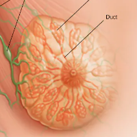
Breast Cancer
Cancer Conditions
Whether you or a loved one are worried about developing breast cancer, have just been diagnosed, are going through breast cancer treatment, or are trying to stay well after treatment, this detailed information can help you find the answers you need. Get the most up-to-date facts about breast cancer, plus tools and resources for every step of your journey. (For information on breast cancer in men, see Breast Cancer in Men .) Breast cancer is a type of cancer that starts in the breast. It can start in one or both breasts. Breast cancer occurs almost entirely in women, but men can get breast cancer , too. Cancer starts when cells begin to grow out of control. (To learn more about how cancers start and spread, see What Is Cancer? ) It’s important to understand that most breast lumps are benign and not cancer (malignant). Non-cancer breast tumors are abnormal growths, but they do not spread outside of the breast. They are not life threatening, but some types of benign breast lumps can increase a woman's risk of getting breast cancer. Any breast lump or change needs to be checked by a health care professional to find out if it is benign or malignant (cancer) and if it might affect your future cancer risk. See Non-cancerous Breast Conditions to learn more. Breast cancers can start from different parts of the breast. The breast is an organ that sits on top of the upper ribs and chest muscles. There is a left and right breast and each one has mainly glands, ducts, and fatty tissue. In women, the breast makes and delivers milk to feed newborns and infants. The amount of fatty tissue in the breast determines the size of each breast. Lobules are the glands that make breast milk. Cancers that start here are called lobular cancers . Ducts are small canals that come out from the lobules and carry the milk to the nipple. This is the most common place for breast cancer to start. Cancers that start here are called ductal cancers . The nipple is the opening in the skin of the breast where the ducts come together and turn into larger ducts so the milk can leave the breast. The nipple is surrounded by slightly darker thicker skin called the areola . A less common type of breast cancer called Paget disease of the breast can start in the nipple. The fat and connective tissue (stroma) surround the ducts and lobules and help keep them in place. A less common type of breast cancer called phyllodes tumor can start in the stroma. Blood vessels and lymph vessels are also found in each breast. Angiosarcoma is a less common type of breast cancer that can start in the lining of these vessels. The lymph system is described below. A small number of cancers start in other tissues in the breast. These cancers are called sarcomas and lymphomas and are not really thought of as breast cancers. To learn more, see Types of Breast Cancer . Breast cancer can spread when the cancer cells get into the blood or lymph system and then are carried to other parts of the body. The lymph (or lymphatic) system is a part of your body's immune system. It is a network of lymph nodes (small, bean-sized glands), ducts or vessels, and organs that work together to collect and carry clear lymph fluid through the body tissues to the blood. The clear lymph fluid inside the lymph vessels contains tissue by-products and waste material, as well as immune system cells. The lymph vessels carry lymph fluid away from the breast. In the case of breast cancer, cancer cells can enter those lymph vessels and start to grow in lymph nodes. Most of the lymph vessels of the breast drain into: Lymph nodes under the arm ( axillary lymph nodes) Lymph nodes inside the chest near the breastbone ( internal mammary lymph nodes) Lymph nodes around the collar bone ( supraclavicular [above the collar bone] and infraclavicular [below the collar bone] lymph nodes) If cancer cells have spread to your lymph nodes, there is a higher chance that the cells could have traveled through the lymph system and spread (metastasized) to other parts of your body. Still, not all women with cancer cells in their lymph nodes develop metastases, and some women with no cancer cells in their lymph nodes might develop metastases later. There are many different types of breast cancer . The type is determined by the specific kind of cells in the breast that are affected. Most breast cancers are carcinomas . The most common breast cancers such as ductal carcinoma in situ (DCIS) and invasive carcinoma are adenocarcinomas , since the cancers start in the gland cells in the milk ducts or the lobules (milk-producing glands). Other kinds of cancers can grow in the breast, like angiosarcoma or sarcoma , but are not considered breast cancer since they start in different cells of the breast. Breast cancers are also classified by certain types of proteins or genes each cancer might make. After a biopsy is done, breast cancer cells are tested for proteins called estrogen receptors and progesterone receptors , and the HER2 gene or protein . The tumor cells are also closely looked at in the lab to find out what grade it is. The specific proteins found and the tumor grade can help decide the stage of the cancer and treatment options. To learn more about the specific tests done on breast cancer cells, see Understanding a Breast Cancer Diagnosis . It’s important to be able to have frank, open discussions with your cancer care team. They want to answer all of your questions so that you can make informed treatment and life decisions. Here are some questions that you can use to help better understand your cancer and your treatment options. Exactly what type of breast cancer do I have? How big is the cancer? Where exactly is it? Has the cancer spread to my lymph nodes or other organs? What is the stage of my cancer? What does it mean? Will I need any other tests before we can decide on treatment? Do I need to see any other doctors or health professionals? What is the hormone receptor status of my cancer? What does this mean? What is the HER2 status of my cancer? What does this mean? What is the grade of my cancer? What does this mean? How do these factors affect my treatment options and long-term outlook (prognosis)? What are my chances of survival, based on my cancer as you see it? Should I think about genetic testing ? What are my testing options? Should I take a home-based genetic test? What would be the reasons for and against testing? How do I get a copy of my pathology report? If I’m worried about the costs and insurance coverage for my diagnosis and treatment, who can help me? How much experience do you have treating this type of cancer? Should I get a second opinion ? How do I do that? Will getting a second opinion delay my treatment and can that affect my outcome? What are my treatment choices? What treatment do you recommend and why? Should I think about taking part in a clinical trial ? What would the goal of the treatment be? How soon do I need to start treatment? How long will treatment last? What will it be like? Where will it be done? Should my biopsy tissue be sent for a gene expression test to help decide if chemotherapy might be helpful for me? Are there other molecular or protein tests that need to be done on my cancer tissue to help decide my treatment options? What should I do to get ready for treatment? What risks or side effects are there to the treatments you suggest? Are there things I can do to reduce these side effects? How will treatment affect my daily activities? Can I still work fulltime? Will I lose my hair? If so, what can I do about it? Will I go through menopause as a result of the treatment? Will I be able to have children after treatment? Would I be able to breastfeed? Do I have time to freeze my eggs before starting treatment? What are my options? What are the chances the cancer will come back (recur) after this treatment? What would we do if the treatment doesn’t work or if the cancer comes back? What if I have transportation problems getting to and from treatment? Is breast-conserving surgery (lumpectomy) an option for me? Why or why not? What are the positive and negative sides of breast-conserving surgery versus mastectomy? How many surgeries like mine have you done? Will you have to take out lymph nodes? If so, would you advise a sentinel lymph node biopsy? Why or why not? What side effects might lymph node removal cause? How long will I be in the hospital? Will I have stitches or staples at the surgery site? Will there be a drain (tube) coming out of the site? How do I care for the surgery site? Will I need someone to help me? What will my breasts look and feel like after my surgery? Will I have normal feeling in them? What will the scar look like? Is breast reconstruction surgery an option if I want it? What would it mean in my case? Can I have reconstruction at the same time as the surgery to remove the cancer? What are the reasons for and against having it done right away or waiting until later? What types of reconstruction might be options for me? Could you recommend a plastic surgeon I could speak to about reconstruction options? Will I need a breast form (prosthesis), and if so, where can I get one? Do I need to stop taking any medications or supplements before surgery? When should I call your office if I’m having side effects or concerns? Once treatment begins, you’ll need to know what to expect and what to look for. Not all of these questions may apply to you, but asking the ones that do may be helpful. How will we know if the treatment is working? Is there anything I can do to help manage side effects? What symptoms or side effects should I tell you about right away? How can I reach you on nights, holidays, or weekends? Will I need to change what I eat during treatment? Are there any limits on what I can do? Can I exercise during treatment? If so, what kind of exercise should I do, and how often? Can you suggest a mental health professional I can see if I start to feel overwhelmed, depressed, or distressed? Will I need special tests, such as imaging scans or blood tests during treatment? If so, how often? Will I need a special diet after treatment? Are there any limits on what I can do? Am I at risk for lymphedema ? What can I do to reduce my risk for lymphedema? What should I do if I notice swelling in my arm? What other symptoms should I watch for? What kind of exercise should I do now? What type of follow-up will I need after treatment? How often will I need to have follow-up exams, blood tests, or imaging tests? How will we know if the cancer has come back? What should I watch for? What will my options be if the cancer comes back? It’s important to be able to have frank, open discussions with your cancer care team. They want to answer all of your questions, so that you can make informed treatment and life decisions. Not all of these questions will apply to you, but they should help get you started. Be sure to write down some questions of your own. For instance, you might want more information about recovery times or you may want to ask about nearby or online support groups where you can talk with other women going through similar situations. You may also want to ask if you qualify for any clinical trials . Don’t be afraid to take notes and tell the doctors or nurses when you don’t understand what they’re saying. You might want to bring another person with you when you see your doctor and have them take notes to help you remember what was said. Keep in mind that doctors aren’t the only ones who can give you information. Other health care professionals, such as nurses and social workers, can answer some of your questions. To find out more about speaking with your health care team, see The Doctor-Patient Relationship . About Breast Cancer What Is Breast Cancer? What Causes Breast Cancer? Types of Breast Cancer Key Statistics for Breast Cancer What’s New in Breast Cancer Research? Breast Cancer Risk and Prevention Breast Cancer Early Detection and Diagnosis Understanding a Breast Cancer Diagnosis Treating Breast Cancer Breast Reconstruction Surgery Living as a Breast Cancer Survivor Non-cancerous Breast Conditions Breast Cancer Videos Infographic: 7 Things to Know About Getting a Mammogram Breast Cancer Quiz Frequently Asked Questions About the American Cancer Society’s Breast Cancer Screening Guideline Breast Cancer Grades Breast Cancer Ploidy and Cell Proliferation Breast Cancer Hormone Receptor Status Breast Cancer HER2 Status Breast Cancer Gene Expression Tests Other Breast Cancer Gene, Protein, and Blood Tests Understanding Your Pathology Report Imaging Tests to Find Out if Breast Cancer Has Spread Breast Cancer Stages Breast Cancer Survival Rates Breast Reconstruction Surgery Living as a Breast Cancer Survivor Source: https://www.cancer.org/cancer/types/breast-cancer.html
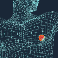
Breast Cancer in Men
Cancer Conditions
Breast cancer occurs mainly in women, but men can get it, too. Many people do not realize that men have breast tissue and that they can develop breast cancer. Cells in nearly any part of the body can become cancer and can spread to other areas. Breast cancer starts when cells in the breast begin to grow out of control. These cells usually form a tumor that can often be seen on an x-ray or felt as a lump. The tumor is malignant (cancer) if the cells can grow into (invade) surrounding tissues or spread (metastasize) to distant areas of the body. To learn more about how cancers start and spread, see What Is Cancer? Until puberty (on average around age 9 or 10), young boys and girls have a small amount of breast tissue consisting of a few ducts located under the nipple and areola (area around the nipple). At puberty, a girl's ovaries make female hormones, causing breast ducts to grow and lobules to form at the ends of ducts. Even after puberty, boys and men normally have low levels of female hormones, and breast tissue doesn’t grow much. Men's breast tissue has ducts, but only a few if any lobules. Breast cancers can start from different parts of the breast. Most breast cancers begin in the ducts that carry milk to the nipple (ductal cancers). Some start in the glands that make breast milk (lobular cancers). Men have these ducts and glands, too, even though they aren't normally functional. There are also types of breast cancer that start in other types of breast cells, but these are less common. A small number of cancers start in other tissues in the breast. These cancers are called sarcomas and lymphomas and are not really thought of as breast cancers. Although many types of breast cancer can cause a lump in the breast, not all do. There are other symptoms of breast cancer you should watch for and report to a health care provider. It’s also important to understand that most breast lumps are benign and not cancer (malignant). Benign breast tumors are abnormal growths, but they do not spread outside of the breast and they are not life threatening. Any breast lump or change needs to be checked by a health care provider to determine whether it is benign or malignant (cancer) and whether it might impact your future cancer risk. Breast cancer can spread when the cancer cells get into the blood or lymph system and are carried to other parts of the body. The lymph system is a network of lymph (or lymphatic) vessels found throughout the body. The lymph vessels carry lymph fluid and connect lymph nodes. Lymph nodes are small, bean-shaped collections of immune system cells. Lymph vessels are like small veins, except that they carry a clear fluid called lymph (instead of blood) away from the breast. Lymph contains tissue fluid and waste products, as well as immune system cells. Breast cancer cells can enter lymph vessels and start to grow in lymph nodes. Most of the lymph vessels of the breast drain into: Lymph nodes under the arm (axillary nodes) Lymph nodes around the collar bone (supraclavicular [above the collar bone] and infraclavicular [below the collar bone] lymph nodes) Lymph nodes inside the chest near the breast bone (internal mammary lymph nodes) If the cancer cells have spread to your lymph nodes, there is a higher chance that the cells could have also traveled through the lymph system and spread (metastasized) to other parts of your body. The more lymph nodes with breast cancer cells, the more likely it is that the cancer may be found in other organs. Because of this, finding cancer in one or more lymph nodes often affects your treatment plan. Usually, surgery to remove one or more lymph nodes will be needed to know whether the cancer has spread. Still, not all men with cancer cells in their lymph nodes develop metastases to other areas, and some men can have no cancer cells in their lymph nodes and later develop metastases. Men can also have some benign (not cancerous) breast disorders. Gynecomastia is the most common male breast disorder. It is not a tumor but rather an increase in the amount of a man's breast tissue. Usually, men have too little breast tissue to be felt or noticed. Gynecomastia can appear as a button-like or disk-like growth under the nipple and areola (the dark circle around the nipple), which can be felt and sometimes seen. Some men have more severe gynecomastia and they may appear to have small breasts. Although gynecomastia is much more common than breast cancer in men, both can be felt as a growth under the nipple, which is why it's important to have any such lumps checked by your doctor. Gynecomastia is common among teenage boys because the balance of hormones in the body changes during adolescence. It is also common in older men due to changes in their hormone balance. In rare cases, gynecomastia occurs because tumors or diseases of certain endocrine (hormone-producing) glands cause a man's body to make more estrogen (the main female hormone). Men's glands normally make some estrogen, but not enough to cause breast growth. Diseases of the liver, which is an important organ in male and female hormone metabolism, can change a man's hormone balance and lead to gynecomastia. Having excess body weight or obesity can also cause higher levels of estrogen in men. Some medicines can cause gynecomastia. These include some drugs used to treat ulcers and heartburn, high blood pressure, heart failure, and psychiatric conditions. Men with gynecomastia should ask their doctors if any medicines they are taking might be causing this condition. Klinefelter syndrome, a rare genetic condition, can lead to gynecomastia as well as increase a man's risk of developing breast cancer. This condition is discussed further in Risk Factors for Breast Cancer in Men . There are many types of benign breast tumors (abnormal lumps or masses of tissue), such as papillomas and fibroadenomas. Benign tumors do not spread outside the breast and are not life threatening. Benign breast tumors are common in women but are very rare in men. The most common types of breast cancer are ductal carcinoma in situ, invasive ductal carcinoma, and invasive lobular carcinoma. Most breast cancers are carcinomas . In fact, breast cancers are often a type of carcinoma called adenocarcinoma , which starts in cells that make glands (glandular tissue). Breast adenocarcinomas start in the ducts (the milk ducts) or the lobules (milk-producing glands). There are other, less common, types of breast cancers, too, such as sarcomas , phyllodes, Paget’s disease and angiosarcomas which start in the cells of the muscle, fat, or connective tissue. Sometimes a single breast tumor can be a combination of different types. And in some very rare types of breast cancer, the cancer cells may not form a lump or tumor at all. When a biopsy is done to find out the specific type of breast cancer, the pathologist also will say if the cancer has spread in to the surrounding tissues. The name of the breast cancer type will change depending on the extent of the cancer. In situ breast cancers have not spread. Invasive or infiltrating cancers have spread (invaded) into the surrounding breast tissue. These general kinds of breast cancer can be further described with the terms outlined above. Ductal carcinoma in situ (DCIS; also known as intraductal carcinoma) is considered non-invasive or pre-invasive breast cancer. In DCIS (also known as intraductal carcinoma), cells that lined the ducts have changed to look like cancer cells. The difference between DCIS and invasive cancer is that the cells have not spread (invaded) through the walls of the ducts into the surrounding tissue of the breast (or spread outside the breast). DCIS is considered a pre-cancer because some cases can go on to become invasive cancers. Right now, though, there is no good way to know for certain which cases will go on to become invasive cancers and which ones won’t. DCIS accounts for about 1 in 10 cases of breast cancer in men. It is almost always curable with surgery. Lobular carcinoma in situ (LCIS) may also be called lobular neoplasia. In LCIS, cells that look like cancer cells are growing in the lobules of the milk-producing glands of the breast, but they haven’t grown through the wall of the lobules. LCIS is not a true pre-invasive cancer because it does not turn into an invasive cancer if left untreated, but it is linked to an increased risk of invasive cancer in both breasts. LCIS is rarely, if ever seen in men. This is the most common type of breast cancer. Invasive (or infiltrating) ductal carcinoma (IDC) starts in a milk duct of the breast, breaks through the wall of the duct, and grows into the fatty tissue of the breast. Once it breaks through the wall of the duct, it has the potential to spread to other parts of the body. Invasive (or infiltrating) ductal carcinoma (IDC) starts in a milk duct of the breast, breaks through the wall of the duct, and grows into the fatty tissue of the breast. At this point, it may be able to spread (metastasize) to other parts of the body through the lymphatic system and bloodstream. At least 8 out of 10 male breast cancers are IDCs (alone or mixed with other types of invasive or in situ breast cancer). Because the male breast is much smaller than the female breast, all male breast cancers start relatively close to the nipple, so they are more likely to spread to the nipple. This is different from Paget disease as described below. Invasive lobular carcinoma (ILC) starts in the milk-producing glands (lobules). Like IDC, it can spread to other parts of the breast and body. ILC is very rare in men, accounting for only about 2% of male breast cancers. This is because men do not usually have much lobular (glandular) breast tissue. This type of breast cancer starts in the breast ducts and spreads to the nipple. It may also spread to the areola (the dark circle around the nipple). The skin of the nipple usually appears crusted, scaly, and red, with areas of itching, oozing, burning, or bleeding. There may also be an underlying lump in the breast. Paget disease may be associated with DCIS or with infiltrating ductal carcinoma. It is rare and accounts for about 1-3% of female breast cancers and a higher percentage (5%) of male breast cancers. Inflammatory breast cancer is an aggressive, but rare type of breast cancer. It makes the breast swollen, red, warm and tender rather than forming a lump. It can be mistaken for an infection of the breast. This is very rare in men. See Inflammatory Breast Cancer for more information. There are some special types of breast cancer that are sub-types of invasive carcinoma. They are much less common than the breast cancers named above. Some of these may have a better or worse prognosis than standard infiltrating ductal carcinoma. Adenoid cystic (or adenocystic) carcinoma Low-grade adenosquamous carcinoma (this is a type of metaplastic carcinoma) Medullary carcinoma Mucinous (or colloid) carcinoma Papillary carcinoma Tubular carcinoma Metaplastic carcinoma (including spindle cell and squamous, except low grade adenosquamous carcinoma) Micropapillary carcinoma Mixed carcinoma (has features of both invasive ductal and lobular) In general, these sub-types are still treated like standard infiltrating carcinoma. It’s important to be able to have frank, open discussions with your cancer care team. They want to answer all of your questions, so that you can make informed treatment and life decisions. Here are some questions that you can use to help better understand your cancer and your treatment options. Don’t be afraid to take notes and tell the doctors or nurses when you don’t understand what they’re saying. You might want to bring another person with you when you see your doctor and have them take notes to help you remember what was said. Not all of these questions will apply to you, but they should help get you started. Be sure to write down some questions of your own. For instance, you might want more information about recovery times or you may want to ask about nearby or online support groups where you can talk with other men going through similar situations. You may also want to ask if you qualify for any clinical trials . Keep in mind that doctors aren’t the only ones who can give you information. Other health care professionals, such as nurses and social workers, can answer some of your questions. To find out more about speaking with your health care team, see The Doctor-Patient Relationship . Exactly what type of breast cancer do I have? How big is the cancer? Where exactly is it? Has the cancer spread to my lymph nodes or other organs? What’s the stage of the cancer? What does that mean? Will I need any other tests before we can decide on treatment? Do I need to see any other doctors or health professionals? What is the hormone receptor status of my cancer? What does this mean? What is the HER2 status of my cancer? What does this mean? How do these factors affect my treatment options and long-term outlook (prognosis)? What are my chances of survival, based on my cancer as you see it? Should I think about genetic testing? What would the pros and cons of testing be? How do I get a copy of my pathology report? If I’m concerned about the costs and insurance coverage for my diagnosis and treatment, who can help me? What treatments are appropriate for me? What do you recommend? Why? How long will treatment last? What will it involve? Where will it be done? What risks or side effects should I expect? Should I think about taking part in a clinical trial? What should I do to get ready for treatment? How much experience do you have treating this type of cancer? Should I get a second opinion ? How do I do that? What would the goal of the treatment be? How soon do I need to start treatment? How will treatment affect my daily activities? Can I still work fulltime? Will I lose my hair? If so, what can I do about it? What are the chances the cancer will come back (recur) after this treatment? What would we do if the treatment doesn’t work or if the cancer comes back? What if I have transportation problems getting to and from treatment? Is breast-conserving surgery (lumpectomy) an option for me? Why or why not? What are the pros and cons of breast-conserving surgery versus mastectomy? How many surgeries like mine have you done? Will you have to take out lymph nodes? If so, would you advise a sentinel lymph node biopsy? Why or why not? What side effects might lymph node removal cause? How long will I be in the hospital? Will I have stitches or staples at the surgery site? Will there be a drain (tube) coming out of the site? How do I care for the surgery site? Will I need someone to help me? What will the scar look like? Do I need to stop taking any medications or supplements before surgery? When should I call your office if I’m having side effects? Once treatment begins, you’ll need to know what to expect and what to look for. Not all of these questions may apply to you, but asking the ones that do may be helpful. How will we know if the treatment is working? Is there anything I can do to help manage side effects? What symptoms or side effects should I tell you about right away? How can I reach you on nights, holidays, or weekends? Will I need to change what I eat during treatment? Are there any limits on what I can do? Can I exercise during treatment? If so, what kind of exercise should I do, and how often? Can you suggest a mental health professional I can see if I start to feel overwhelmed, depressed, or distressed? Will I need special tests, such as imaging scans or blood tests? How often? What are the chances my cancer might come back? What will we do if that happens? What type of follow-up will I need after treatment? Will I need a special diet after treatment? Are there any limits on what I can do? Am I at risk for lymphedema? What can I do to reduce my risk for lymphedema? What should I do if I notice swelling in my arm? What other symptoms should I watch for?What kind of exercise should I do now? What type of follow-up will I need after treatment? How often will I need to have follow-up exams, blood tests, or imaging tests? How will we know if the cancer has come back? What should I watch for? About Breast Cancer in Men What Is Breast Cancer in Men? Key Statistics for Breast Cancer in Men What's New in Research for Breast Cancer in Men? Causes, Risk Factors, and Prevention Early Detection, Diagnosis, and Staging Treating Breast Cancer in Men After Treatment If You're a Man with Breast Cancer Surgery for Breast Cancer in Men Radiation Therapy for Breast Cancer in Men Chemotherapy for Breast Cancer in Men Hormone Therapy for Breast Cancer in Men Targeted Drug Therapy for Breast Cancer in Men Immunotherapy for Breast Cancer in Men Treatment of Breast Cancer in Men, by Stage Health Professionals Who Are Part of a Cancer Care Team Source: https://www.cancer.org/cancer/types/breast-cancer-in-men/about.html
Can Cancer Be Cured? Remission, Survival and Survivorship
Cancer Conditions
After a cancer diagnosis, many people want to know whether treatment will get rid of the cancer, how long they are likely to live, and what life will be like now. This topic explains what doctors mean by cure, remission, survival statistics and cancer survivor. A cure means no trace of the cancer is left after treatment — sometimes called NED, "no evidence of disease" — and the cancer is not expected to come back. But no doctor can be sure a cancer will never return, and it usually takes time to know. The longer you stay cancer-free, the better the chance it will not return. Cancer that comes back is called a recurrence. Most recurrences happen within the first 5 years, but later ones are possible, and some doctors call a cancer cured if it has not returned by 5 years. "Cure" is a hard word to use with cancer, because a few cancer cells can remain after tests show none, and they can cause a return weeks, months or years later. When tests cannot find cancer, doctors more often say it is "in remission" rather than "cured." Remission is a time when the cancer is responding to treatment or is under control — it does not always mean cured. In complete remission , all signs of cancer are gone and tests find no cancer cells. In partial remission , the cancer shrinks but does not go away. A remission can last from weeks to years, and treatment may or may not continue during it, depending on the cancer. If the cancer returns, more treatment may bring another remission. Statistics describe what happened to large groups of people with the same diagnosis. They cannot predict what will happen to one person, but they can give an idea of what to expect. Survival rate — the percentage of people alive at a set time after diagnosis. Overall survival rate — the percentage of people with a given type and stage of cancer who are alive at a set time after diagnosis. Cancer-specific survival rate — the percentage of people with a given type and stage who have not died from their cancer at a set time after diagnosis. 5-year relative survival rate — the percentage alive 5 years after diagnosis, leaving out people who die of other causes. Survival rates can cover any length of time, but researchers and doctors usually look at 5-year relative survival. The American Cancer Society calls anyone who has ever been diagnosed with cancer a cancer survivor, wherever they are in their illness. Survivorship looks different for each person. You may be cancer-free but live with late or long-term side effects. You may go into complete remission and later need treatment again for a recurrence. Or you may live with cancer as a chronic condition and keep having treatment to control it. Everyone diagnosed with cancer has their own long-term needs, and taking steps to live well after cancer can help you feel better and improve your long-term health. Not everyone likes the word "survivor," and each person has the right to describe their own experience. Is the goal of my treatment to cure my cancer, or to control it? If tests find no cancer after treatment, will you call it a cure or a remission, and why? What is the 5-year relative survival rate for my type and stage, and how well does it apply to me? How likely is my cancer to come back, and how will you watch for a recurrence? Which late or long-term side effects should I watch for after treatment? Source: https://www.cancer.org/cancer/understanding-cancer/can-cancer-be-cured.html
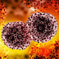
Cancer
Cancer Conditions
1 in 2 men and 1 in 3 women will be diagnosed with cancer in their lifetime. If you’ve been recently diagnosed, or know someone who has, we’re here with answers, guidance, and support. Finding out you have cancer can bring many changes for you and your loved ones. Here, we answer many questions about cancer and making treatment decisions. We also tell you what you can expect from the people and services that are there to help you cope with cancer. This information is not meant to replace the advice of your health care provider. Please keep in mind every patient's situation is different. Because of this, there are questions you will need to ask your doctor. Talking with them is the best way to understand what’s going on with your body, what you can expect, and how treatment will help. AFTER DIAGNOSIS: A GUIDE FOR PATIENTS AND FAMILIES [PDF BOOKLET] Cancer can start any place in the body. It starts when cells grow out of control and crowd out normal cells. This makes it hard for the body to work the way it should. What Is Cancer? Is Cancer Contagious? Can Cancer Be Cured? How to Find Cancer Information on the Internet Cancer Glossary The History of Cancer Learn about the tests used to diagnose and stage cancer, and get help understanding your test results. Signs and Symptoms of Cancer Exams and Tests for Cancer Lymph Nodes and Cancer Cancer Staging Advanced cancers have usually spread from where they started to other parts of the body. This is known as metastatic cancer. But not all advanced cancers are metastatic. Advanced and Metastatic Cancer Understanding Recurrence Before getting any treatment, your doctor will first need to learn more about your cancer. The results of a biopsy, blood tests, physical exams, and imaging tests (x-rays or scans), along with any problems the cancer is causing are used to decide which treatment options might be best for you. Your doctor may also talk with other experts to get their opinions. Your doctor will then discuss the goal of treatment and the pros and cons of each treatment option with you. Making Decisions and Managing Your Treatment Choosing Your Treatment Team Questions to Ask Your Doctor Treatment Types It can be hard to talk about cancer, even with the people you love. Your loved ones may also have a hard time talking about cancer. It’s not easy for them to know what to say to help you or make you feel better. Here are some tips to help you and your loved ones deal with cancer. Telling Others About Your Cancer Helping Children When a Family Member or Someone They Know Has Cancer At first, most people need some time to adjust to the fact that they have cancer. It’s normal to feel disbelief, shock, fear, and anger. You may need some time to absorb and understand what your diagnosis and treatment options mean for you and your loved ones. Coping and Living Well During Cancer Treatment A cancer diagnosis affects close friends and family too. Find out what to expect if you become a caregiver for a person with cancer, and get tips for making sure that you take care of yourself as well. READ MORE This online, interactive guide can empower caregivers with the resources and information they need – from anywhere and at any time. It can help you: Learn how to care for yourself as a caregiver. Better understand what your loved one is going through. Develop skills for coping and caring. Take steps to help protect your health and well-being. This guide is also available as a downloadable PDF . READ MORE This video series offers educational support as you assist with the everyday needs of your loved one, as well as self-care techniques to improve your own quality of life. Physical Care Training: drain care, lifting, pain management, medication management, symptom/side effect management, and identifying signs of infection. Caregiver Self-Care: nutrition, physical activity, stress management and coping, dealing with fear of recurrence, and deep breathing/meditation. Advocacy: how to effectively communicate your loved one’s needs to the cancer care team. Caregiver Stories: caregivers share their experiences through the ups and downs of their journey. This series is also available in Spanish . Here you'll find information on what to expect if you are caring for someone with cancer, and find out what you need to know when caring for a loved one at home. What a Cancer Caregiver Does Family and Medical Leave Act (FMLA) Managing Health Insurance Issues Managing Side Effects Caring for someone who is sick, taking on new responsibilities, and worrying about the future can be exhausting at the very least. Here you can find tips on making sure you also take care of yourself. You'll also find checklists that can help caregivers spot signs of anxiety or depression in yourself or others. If You’re About to Become a Cancer Caregiver Being a Caregiver [PDF] If someone close to you has cancer, do you find it hard to talk to them about it? Do you worry that you'll say the wrong thing? If so, you're not alone. Find out how to be supportive and helpful when you talk with someone who has cancer. How to Be a Friend to Someone with Cancer When Someone You Know Has Cancer READ MORE If your child or teenager has cancer, go here to get information about what to expect. You can find information on coping with the diagnosis, dealing with financial and insurance issues, and nutrition for children with cancer, as well as detailed information about many cancers that affect children and teens. READ MORE The American Cancer Society has programs and services to help people with cancer and their loved ones understand cancer, manage their lives through treatment and recovery, and find the emotional support they need. And best of all, our help is free. If you’re facing a cancer diagnosis, learning more and connecting with others can be a source of support and comfort. Visit these resources from the American Cancer Society and our partners to find what’s right for you. https://www.cancer.org/support-programs-and-services/online-communities.html In this section you'll find general information about the types of treatments used against cancer. This includes both traditional therapies (such as surgery, chemotherapy, and radiation therapy), newer forms of treatment (including information on clinical trials), and complementary and alternative therapies. We'll also discuss the possible side effects of these treatments. If you or someone you care for is facing treatment for a new cancer diagnosis or cancer that has come back, you probably have lots of questions that need answered. Learn about how to approach making decisions about treatment, and what to ask your health care team. Making Decisions and Managing Your Treatment Choosing Your Treatment Team Planning cancer treatment can take time. Find out what you need to know about the most common types of cancer treatment, such as surgery, chemotherapy, radiation therapy, and immunotherapy. Learn how they work and what to expect if they are part of your treatment plan. Treatment Types Clinical Trials How to Interpret News About New Cancer Treatments Cancer Treatment Videos Get information about the physical side effects that can be caused by different cancer treatments and what you can do to manage them. Managing Cancer-related Side Effects Palliative Care Find your cancer type for more specific information on how a certain type of cancer is treated, or see the latest news in cancer treatment. Find Your Cancer Type Latest Treatment News The American Cancer Society offers programs and services to help you during and after cancer treatment. Below are some of the resources we provide. We can also help you find other free or low-cost resources available. 24/7 Cancer Helpline Our 24/7 cancer helpline provides support for people dealing with cancer. We can connect you with trained cancer information specialists who will answer questions about a cancer diagnosis and provide guidance and a compassionate ear. Call us at 1-800-227-2345 Schedule a video chat For cancer patients, getting the right treatment sometimes requires traveling away from home. Lodging expenses can present a significant financial barrier to receiving lifesaving treatment, and financial barriers are known to contribute to disparities in cancer outcomes. We help remove that financial barrier through our Hope Lodge program, lodging grants to local healthcare system partners, and a partnership with Extended Stay America. Find a Hope Lodge Become a Hope Lodge Volunteer Extended Stay America If transportation to treatment is a concern, we may be able to help. In some communities, our Road To Recovery volunteer drivers provide rides to patients who have no way to get to their cancer treatment. We also provide transportation grants to local healthcare systems partners so they can provide transportation assistance to their patients. Get a ride to cancer treatment Become a Road To Recovery volunteer Road To Recovery Rider Guide The Cancer Survivors Network provides a safe online connection where cancer patients and caregivers can find others with similar experiences. You can participate on discussion boards, join a chat room, and build your own support network. Visit the Cancer Survivors Network If you have breast cancer, you may want to connect with someone who knows what you’re feeling – someone who has “been there.” Through our Reach To Recovery website and app, you can match with a trained volunteer who is a breast cancer survivor. Get support for breast cancer Become a Reach To Recovery volunteer Cancer and cancer treatment can alter a patient’s appearance. Our “tlc” Tender Loving Care program offers affordable wigs, hats, scarves, and mastectomy products. Shop “tlc“ SEARCH FOR PROGRAMS IN YOUR AREA You don’t have to face a cancer diagnosis alone. Talk or chat live with a trained cancer information specialist or find support in your own community. Call us: 1-800-227-2345 Source: https://www.cancer.org/
Cancer Glossary
Cancer Conditions
This glossary explains 748 words that you can see in cancer reports, test results and conversations with your care team. Each entry gives the word, how to say it (when a pronunciation is available) and what it means in plain language. "See also" points to a related entry in this glossary. Words are in alphabetical order. No entries start with J, Y, Z or a number. A definition tells you what a word means in general. It does not tell you what it means for you. Ask your doctor or nurse to explain any word in your own report. For the main ideas behind these words, see What Is Cancer? , Genes and Cancer and Can Cancer Be Cured? Can you explain the words in my pathology report in plain language? What is the exact name of my cancer, and what is its stage or grade? Which words in my test results are the most important for my treatment? Can you write down the names of my treatments and medicines for me? Where can I find reliable explanations of the terms you use? A · B · C · D · E · F · G · H · I · K · L · M · N · O · P · Q · R · S · T · U · V · W · X abdomen [AB-duh-men]: The belly area, below the chest and above the pelvis. Organs here include the stomach and the lowest end of the esophagus, the small and large intestines, the liver, the gallbladder, the spleen, the pancreas and others. ablate [ab-late]: To destroy an organ or body tissue, or to stop it from working. See also: ablation. ablation [a-BLAY-shun]: Destroying or taking out all or part of a tissue or body part, or making an organ stop working. Surgery is one way. Drugs are another: for example, medicines that stop the ovaries or testicles from making hormones count as ablation, the same as removing those organs. Heat, freezing and chemicals can also ablate tissue and tumors. Also called: ablative (ab-LAY-tive) therapy. abscess [ab-SES]: Pus that has collected in a tissue, an organ or another part of the body. absolute risk [ab-so-loot risk]: The chance that a person gets a disease, such as a type of cancer, within a set period of time. It is given as a fraction or a percentage. For example, a man in the US has an average lifetime absolute risk of colorectal cancer of about 4%. This means that about 4 men in every 100 will get colorectal cancer during their lives. acquired mutation [uh-KWY-erd myoo-TAY-shun]: A change in DNA that starts in one body cell after birth. Every new cell that comes from that cell has the same change. Sometimes these changes can lead to cancer. Inherited mutations are different: they are present from birth in almost all cells. Parents do not pass acquired mutations to their children. actinic keratosis [ak-TIH-nik KAYR-uh-TOH-sis]: A rough, raised patch of skin that can form after much time in the sun. These patches are not cancer, but over time a small number can turn into squamous cell cancer. They often occur on skin that gets a lot of sun, such as the face, scalp, neck, forearms and backs of the hands. Also called: solar keratosis. active surveillance [AK-tiv ser-VAY-lents]: Close watching of a patient's condition with no treatment. The patient has certain exams and tests at regular times. If the results change, the doctor may recommend treatment. This approach can be part of the plan for some cancers, such as prostate cancer. adenocarcinoma [A-deh-noh-KAR-sih-NOH-muh]: A cancer of glandular tissue. Glands make a substance and release it. Two examples are breast tissue, which makes milk, and the gland cells of the prostate, which make seminal fluid. adenoma [A-deh-NOH-muh]: A non-cancerous growth of glandular tissue, the tissue that produces a substance. Examples: in the breast, the ducts and lobules produce milk; in the prostate, gland cells produce seminal fluid. adenomatous polyp [ad-no-muh-tus or ad-uh-NO-muh-tus pa-lip]: A growth made of abnormal gland cells. It is not cancer, but it can turn into cancer, so doctors usually take it out when they find it. The colon can have 3 kinds of adenoma: tubular, villous and tubulovillous. Their cells have different arrangements. The kinds overlap, so a single adenoma can show features of both the tubular and the villous kind. Related terms: polyp, glandular tissue. adhesions [ad-HEE-zhunz]: Scar tissue that can form after surgery or an injury. When this tissue tightens, it can join organs that are normally separate. Sometimes this causes problems, such as a partial or full blockage of the intestine. adjuvant therapy [ad-juh-vunt]: Treatment given in addition to the main treatment. Usually it means hormone therapy, chemotherapy, radiation therapy or immunotherapy given after surgery, to improve the chance of a cure or to keep the disease under control. Compare to: neoadjuvant therapy. advanced cancer : Cancer that has moved beyond the primary site, where it started. If it has grown past its starting body part but has not reached other parts of the body, doctors call it locally advanced. If it has reached other parts of the body, they call it metastatic. advance directive : A legal paper in which a person states, ahead of time, the health care they want if they lose the ability to decide. Their providers and family can read it. It can cover whether to start life-sustaining treatment, or when to stop it. A second kind names a person who will decide for them if they cannot. aggressive cancer : Cancer that forms, grows or spreads fast. AJCC Staging System : The staging system of the American Joint Committee on Cancer. It uses the TNM system to show how much cancer there is and where it is in the body. T gives the size and location of the tumor and whether it has grown into nearby areas. N tells whether the cancer has spread to nearby lymph nodes. M tells whether the cancer has metastasized (spread to distant parts of the body). allele : One version of a gene. For a given trait, a person gets one allele from each parent, so most people have 2 copies of each gene. Some alleles are dominant and some are recessive. Dominant allele: one copy is enough to give the dominant trait. Recessive allele: two copies are needed to give the recessive trait. allogeneic stem cell transplant [A-loh-jeh-NAY-ik]: A transplant of a donor's bone marrow or stem cells. The donor's tissue type must closely match the patient's. The donor can be a relative or a stranger. The new cells take the place of blood-forming cells that disease or cancer treatment destroyed. See also: stem cells. alopecia [al-o-PEE-shuh]: Loss of hair. It can affect the scalp, body, eyebrows, eyelashes and pubic hair. It often occurs with chemotherapy or radiation therapy. In most people, the hair grows back after treatment stops. alpha blocker [al-fuh]: A drug that relaxes smooth muscle. Doctors sometimes give it to men who have difficulty passing urine because of benign prostatic hyperplasia (BPH). It improves the flow of urine and blood. alternative medicine : Treatment that is unproven or shown not to work, used in place of standard, proven care. Examples are special diets, some herbs or supplements and very high doses of vitamins. The American Cancer Society advises talking with your cancer care team before you try one. Some can cause serious or even fatal side effects. With others, the biggest danger is missing the benefit of standard treatment. See also: complementary therapy. ambulatory [AM-byou-luh-tor-ee]: Walking, or able to walk. An ambulatory care center treats outpatients, who go home the same day and do not stay in a hospital overnight. These centers often do short treatments and procedures. Amsterdam criteria [am-STIR-dam cry-TEER-ee-uh]: Criteria that doctors use to find families that may have hereditary non-polyposis colon cancer (HNPCC). Some people with HNPCC do not meet these criteria. People who do meet them may want to think about genetic testing. analog [an-uh-log]: A substance that is close to another one but not the same. One example is a version of a natural substance that is made in a lab. See also: luteinizing hormone-releasing hormone analogs or LHRH analogs. anastomosis [uh-NAS-toh-MOH-sis]: A procedure that removes a diseased part of a tube-shaped body structure and joins the healthy ends together again. androgen [AN-druh-jen]: Hormones that both males and females have, but at much higher levels in males. People often call them male sex hormones. Testosterone is the main androgen. anecdotal report [an-neck-DOE-tul]: A report about one patient or a few patients, based on personal experience. It does not give a full description, and it is not a scientific study result. anemia [uh-NEE-mee-uh]: A condition in which the red blood cell count is low. anesthesia [an-es-THEE-zhuh]: Loss of feeling caused by medicines or other substances, given so that patients do not feel pain during surgery or other procedures. General anesthesia makes you unconscious (a deep sleep), so you do not feel pain. Local or regional anesthesia makes only one area numb, so you do not feel pain there. anesthesiologist [an-es-thee-zee-AHL-uh-jist]: A doctor who specializes in drugs and other agents, such as gases, that block all feeling, cause deep sleep or relieve pain. Usually this doctor works during surgery. aneuploid [AN-you-ploid]: See: ploidy. angiogenesis [an-jee-o-JEN-uh-sis]: The growth of new blood vessels. Some cancer treatments block angiogenesis. This helps stop blood from reaching ("feeding") the tumor. angiography [an-jee-AH-gruh-fee]: A test that puts contrast dye directly into a blood vessel that leads to the area of interest. Then a series of x-rays shows the doctors the blood vessels around a tumor. angiosarcoma [an-jee-o-sar-KO-muh]: Cancer that starts in the cells that line blood vessels or lymph vessels. anorexia [an-uh-REK-see-uh]: Loss of appetite. The cancer itself or treatments such as chemotherapy can cause it. anterior [an-teer-ee-yer]: At or close to the front. anti-androgens [an-tee-AN-druh-jens or an-tie-AN-druh-jens]: Drugs that stop the body from using androgens (male hormones). They are pills that are taken up to 3 times each day. To help treat prostate cancer, they are usually given together with orchiectomy (surgery) or LHRH analogs (medicines). anti-angiogenesis agent [an-tee-an-jee-o-JEN-uh-sis or an-tie-an-jee-o-JEN-uh-sis]: A drug that stops a tumor from making blood vessels, so the tumor loses its blood supply. antibiotic [an-tee-by-AH-tick or an-tie-by-AH-tick]: A drug that treats infections caused by bacteria and other germs. antibody [AN-tih-bah-dee]: A protein that immune system cells make and release into the blood. Antibodies protect the body against foreign agents, such as bacteria. The body makes them in response to antigens, which are substances that cause a specific immune response. Each antibody can destroy only one specific antigen. anti-emetic [an-tee-ih-MEH-tik or an-tie-ih-MEH-tik]: A drug that prevents or reduces nausea and vomiting. anti-estrogen [an-tee-ES-tro-jen or an-tie-ES-tro-jen]: A substance, such as the drug tamoxifen, that stops cells from making estrogen. Anti-estrogens treat breast cancers that need estrogen to grow, and researchers are studying them for other cancers. An anti-estrogen is one type of hormone antagonist. Also called: estrogen blocker. antigen [an-tuh-jen]: A foreign substance that triggers the immune system to make antibodies. Bacteria and viruses carry antigens, and the immune response to them helps people fight infection. Some cancer cells carry antigens that lab tests can detect. Doctors use these to diagnose those cancers and to follow how treatment works. Other antigens on cancer cells take part in immune reactions that may help the body fight the cancer. antimetabolites [an-tee-muh-TAB-o-lites or an-tie-muh-TAB-o-lites]: Substances that disrupt chemical processes in the body, for example the making of proteins, DNA (deoxyribonucleic acid) and other chemicals that a cell needs to grow and copy itself. As cancer drugs, they block DNA production. Then the cells cannot divide and the tumor cannot grow. antioxidants [an-tee-OX-uh-dunts or an-tie-OX-uh-dunts]: Substances that protect cells from oxidation damage, which happens during metabolism. Vitamins C and E and beta-carotene are examples. Antioxidants in food are thought to lower the risk of some cancers. But in some people, some antioxidant supplements are linked with a higher cancer risk. APC gene : A gene that helps cells grow at a balanced rate. If a person inherits a change in it, they can get familial adenomatous polyposis (FAP) or Gardner syndrome. A mutation (change) in this gene can cause hundreds of polyps to grow in the colon. apheresis [a-fur-REE-sis]: A procedure that takes blood out of the body, removes one part of it and puts the rest back into the body. It is called plasmapheresis when plasma is removed, leukapheresis when white blood cells are removed and platelet pheresis when platelets are removed. Also called: pheresis (fur-REE-sis). apoptosis [a-pop-TOE-sis]: Programmed cell death. Genes control it and make cells die at certain times, for example when their DNA is damaged. Cancer cells can block this process. Some cancer drugs cause apoptosis. aromatase inhibitors : Drugs that block estrogen production by the adrenal glands. Doctors use them for hormone-sensitive breast cancer in women who have gone through menopause. Examples are anastrozole (Arimidex®), letrozole (Femara®) and exemestane (Aromasin®). Trials are checking whether they can also lower the risk of breast cancer in women after menopause. aspiration [asp-er-A-shun]: To remove a liquid, a gas or pieces of tissue by suction. It can also mean to breathe food or liquid into the lungs by accident (aspirate). asymptomatic [a-simp-tuh-MAT-ik]: Having no signs or symptoms of a disease. ataxia-telangiectasia mutation (ATM) [a-tax-ee-yuh tel-an-jee-eck-TAY-zhuh]: An inherited change in a gene whose job is to repair damaged DNA. Ataxia-telangiectasia itself is a rare childhood disease that is inherited and slowly gets worse. It causes loss of muscle control and a weak immune system, and it raises cancer risk. People who carry the mutation can have a higher risk of several cancers. attributable risk [at-trib-ut-a-ble risk]: How much a known factor adds to the risk of a disease, such as cancer. It is usually a percentage. It compares people who had the risk factor and got the disease with people who did not have the factor but got the same disease. For example, an attributable risk of 80% for a factor and a cancer means that 80% of people with this cancer had the factor and 20% did not. atypia [a-tip-ee-yuh]: Not normal (atypical). The word often describes how cancer cells or precancer cells look. atypical [a-tip-uh-kul]: Not usual; abnormal. autologous [aw-tahl-uh-gus]: From the person's own body. It describes a procedure that uses the patient's own blood or tissue. For example, rebuilding a woman's breast with tissue taken from her own body is autologous tissue construction. axillary dissection [ax-ill-air-ee di-sek-shun]: Surgery that removes the lymph nodes in the armpit (axillary nodes). These nodes are examined with a microscope for cancer. barium sulfate [bear-ee-um sul-fate]: A chalky liquid that coats the digestive tract so it shows up on x-rays. You swallow it for an upper gastrointestinal (GI) series. For a lower GI series, it goes into the rectum as a barium enema. See also: gastrointestinal tract or GI tract. basal cell carcinoma [bay-sul or bay-zul sell car-sin-O-ma]: Skin cancer of the most common type. It starts in the basal cells, which form the deepest layer of the epidermis (the skin's outer layer). It mostly appears on skin that gets sun, often the head and neck. It grows slowly and rarely spreads to distant parts of the body. basic science : Science that gives the knowledge and background that later research on human health problems needs. In cancer research, it is often lab work in fields such as biochemistry, cell biology or genetics. This work does not aim to treat one specific cancer, but later it can be part of the basis for a treatment. Also called: pure science. behavioral research : Research on why people act the way they do. Its results can help people choose healthy lifestyles and follow screening and treatment guidelines that save lives. benign [be-nine]: Not cancer; not malignant. beta carotene [bay-tuh KAIR-uh-teen]: A substance found mostly in yellow and orange fruits and vegetables. It acts as an antioxidant, and the body changes it into vitamin A. Bethesda criteria [beth-ez-duh cry-teer-ree-yuh]: A checklist of traits that are common in people who have hereditary non-polyposis colon cancer (HNPCC). Fitting the checklist does not mean a person has HNPCC; most who fit it do not. Genetic testing for HNPCC is something they may want to consider. bilateral [by-lat-er-ul]: Affecting both sides of the body. Cancer in both breasts, for example, is bilateral breast cancer. Compare to: unilateral. biologic response modifiers [by-o-LA-jick re-spons MOD-uh-fie-urs]: Substances, such as the drug interferon, that make the immune system stronger against cancer. This treatment is sometimes called biologic therapy. biomarkers [BY-o-mar-kers]: Substances, such as genes, proteins and hormones, that tests can look for to learn key facts about a cancer. They can help detect cancer, show whether treatment is working, or watch for a cancer that has returned (recurred). Also called: tumor markers. biomarker testing [BY-o-mar-ker]: Tests for genes, proteins or other substances. They help find out whether: A cancer is more likely to grow and spread A certain treatment is likely or not likely to help Treatment is working A person may have a higher risk of some cancers A person may have a certain type of cancer (but a biopsy is usually needed to be sure) Cancer may be coming back Also called: tumor testing, tumor genetic testing, tumor subtyping, genomic testing or genomic profiling. biopsy [by-op-see]: Taking out a small piece of tissue, or some cells, to check them for cancer. Biopsies come in several types. biopsy gun [by-op-see]: A tool that takes core biopsy samples. It is often used for prostate biopsies. bisphosphonates [bis-FAHS-fun-ates]: Drugs that slow osteoclasts, the cells that break bone down. When cancer reaches the bones, it can make these cells overactive. The drugs can ease pain and lower high calcium levels caused by cancer in the bones. They can also slow that cancer's growth, delay or prevent fractures and, in some people, strengthen bones. blood count : How many cells a measured sample of blood contains. body image : A person's own view of their body, and how they believe other people see it. bone marrow : The soft, spongy tissue in the hollow center of some bones. The body makes new blood cells here. bone marrow aspiration and biopsy [asp-er-A-shun and by-op-see]: A procedure that puts a needle into the center of a bone, usually the hip bone or breast bone. The needle takes out a small amount of bone marrow, which is examined with a microscope. bone marrow transplant : A treatment that gives healthy stem cells to replace the stem cells that high doses of chemotherapy, radiation therapy or both have destroyed. The marrow can come from the patient (autologous) or from a donor (allogeneic). Also called: stem cell transplant. bone scan : An imaging test that gives important information about the bones, such as where cancer may have spread in them. A small amount of radioactive material (radioisotope) goes into a vein. It collects in areas of bone where cancer may be. These areas ("hot spots") show on the pictures. bone survey : A set of x-rays that covers every bone in the body. It may be used to look for cancer that has spread to bone. Also called: skeletal survey. brachytherapy [brake-ee-THER-uh-pee]: Radiation treatment from inside the body. The radiation source is usually sealed in a small holder called an implant, which is put very near the tumor or into it. Compare to: external beam radiation therapy. brain scan : An imaging method that finds anything abnormal in the brain, such as brain cancer or cancer that has spread to the brain from another part of the body. It can be done in an outpatient clinic. The only pain is the needle stick when a radioactive substance (radioisotope) goes into a vein. The substance collects in abnormal areas, and these areas show on the pictures. BRCA1 : A gene that, when it is damaged (mutated), gives a person a higher risk of breast, ovarian, pancreatic, prostate and other cancers than people without the mutation. BRCA2 : A gene that, when it is damaged (mutated), gives a person a higher risk of breast, ovarian, pancreatic, prostate and other cancers than people without the mutation. BRCAPRO : A tool that helps health professionals estimate a woman's breast cancer risk from certain risk factors. breast specialist : A health care professional with a special interest in breast health. This person may have special knowledge, but medical licensing boards do not certify breast care as a specialty. bronchoscopy [brong-kah-skuh-pee]: An exam of the bronchi in the lungs with a thin, flexible tube that has a light. The tube, called a bronchoscope, goes down the throat. CA 19-9 : A tumor marker that colorectal, stomach, bile duct and pancreatic cancers sometimes make. Conditions that are not cancer can also make it. cachexia [ka-kek-see-uh]: A large loss of weight and muscle caused by loss of appetite and poor nutrition. calcifications [kals-if-ik-A-shuns]: Very small deposits of calcium in the breast, alone or in groups. Mammograms often show them. The smallest ones may be called microcalcifications. They show changes in the breast that may need more mammograms or a biopsy. cancer : A group of diseases in which body cells change and start to grow without control. Most cancers make a lump or mass, called a tumor. A tumor can be benign, which means it is not cancer, or malignant, which means it is cancer. A malignant tumor can grow into healthy tissue and destroy it. cancer care team : All the health care professionals who find, treat and look after a person with cancer. They may work together formally or informally. Usually one of them coordinates the team. cancer cell : A cell that divides and multiplies in an abnormal way. It can spread through the body and push out normal cells and tissue. Cancer cells form because of many changes in certain genes. cancer-related fatigue : Unusual tiredness that continues and that can come with cancer or its treatment. It can be very strong, last a long time and get in the way of daily life. Rest does not always make it better. cancer screening tests : See: screening. cancer surveillance : The continuous, timely and organized collection and study of public health information about new cancer cases, how far the disease has spread, screening tests, treatment, survival and cancer deaths. cancer susceptibility genes : Genes that, with certain mutations, can raise a person's risk of some cancers. These genes usually come from one or both parents. cancer vaccine : A vaccine that trains a person's immune system to attack cancer cells. It can help treat a cancer, or help keep it from returning after other treatment. A few vaccines, such as the HPV vaccine, may instead help prevent some cancers. cannula : A narrow tube. Different types can hold tissues open during laparoscopy or give IV medicines and fluids. capsular contracture : Hardness and change of shape in an area, caused by scar tissue that forms around an implant in the body. capsule formation : Scar tissue that can form around an implant because the body reacts to a foreign object. carcinoembryonic antigen (CEA) [car-sin-o-em-bre-ON-ic an-tuh-jin]: A substance that is normally in some tissues of an unborn baby. In an adult, it can be a sign of cancer, mostly cancer that starts in the digestive system. Tests for it can help doctors find out whether colorectal cancer has come back after treatment. carcinogen [car-sin-o-jin]: Any substance that causes cancer or helps it grow. carcinoid syndrome [car-sin-oyd sin-drom]: A group of symptoms caused by carcinoid tumors that release large amounts of hormones. The symptoms include a flushed face, wheezing, diarrhea, a fast heartbeat and others. carcinoid tumor [car-sin-oyd]: A tumor that grows from neuroendocrine (nerve and endocrine) cells. It can release hormones that cause symptoms such as a flushed face, wheezing, diarrhea and a fast heartbeat. carcinoma [car-sin-O-ma]: Cancer that starts in the skin or in the layer of cells that lines organs (epithelial cells). carcinoma in situ [car-sin-O-ma in sy-too]: An early cancer that has stayed in the organ where it started. It has not grown into other parts of that organ, and it has not spread to distant parts of the body. Most can be cured. cardiologist : A doctor who specializes in the heart and blood vessels. People with cancer may see one for heart disease or for heart problems caused by treatment. case manager : The cancer care team member who organizes the patient's care from diagnosis through treatment and recovery. This person often deals with the insurance company and links the patient and family to helpful resources. castration [cass-tray-shun]: Surgery, radiation or medicine that stops the ovaries or testicles from making hormones. catheter [cath-it-ur]: A thin, flexible tube that lets fluids go into or out of the body. For example, a Foley catheter drains urine, and an IV catheter gives fluids or medicines into a vein. causal association or causal link [kaw-zul]: A link in which one factor is believed to bring about a result. For example, smoking is believed to cause lung cancer, so the two have a causal link. CDH1 : A gene. An inherited mutation in it can cause hereditary diffuse gastric cancer, a rare stomach cancer, to start at a young age. In women, changes in this gene also raise the risk of lobular breast cancer. cell : The basic building block of all living things. Cells replace themselves when they split to make new cells (mitosis). In cancer, the processes that control how new cells form and old cells die do not work correctly. cell cycle : The steps a cell goes through before it divides. Some chemotherapy drugs work by blocking a step in this cycle. centimeter [SIN-tuh-me-ter]: A metric unit of length, written cm, equal to 1/100 of a meter. About 2½ cm equal 1 inch. See also: millimeter. cerebrospinal fluid [suh-REE-bro-spy-nuhl]: A clear liquid around the brain and spinal cord that cushions them. Also called: CSF. cGy : See: Gray, under radiation dose. chaplain : A member of the clergy who helps with the spiritual needs of the patient and family. A chaplain can usually help people of many faiths, denominations and beliefs. CHEK2 : A gene that, when it is damaged or mutated, can raise the risk of breast cancer and other cancers. A person can inherit a damaged CHEK2 gene. chemo brain [key-mo brain]: A change in thinking. A person may feel less mentally "sharp," forget some things, or have difficulty finishing tasks, concentrating or learning new skills. It can occur at any time with cancer, before or after treatment. chemoprevention [key-mo-pre-VEN-shun]: Using drugs, chemicals or other substances to try to prevent cancer, or to lower the chance that cancer comes back. chemotherapy [key-mo-THER-uh-pee]: Medicines that treat cancer. You can get them alone or together with surgery, radiation therapy, hormone therapy, targeted therapy or immunotherapy. They can go into a vein as an infusion, be given as a shot or be swallowed. Often called: chemo. chromosome [KROM-uh-som]: A long strand of DNA that holds genes, the basic units of heredity. Each chromosome can hold hundreds or thousands of genes. Most human cells have 23 pairs. In each pair, one comes from the mother and one from the father. chronic inflammatory bowel disease : See: inflammatory bowel disease (IBD). chronic obstructive pulmonary disease (COPD) [kron-ick ob-struck-tiv PULL-muh-nerr-ee]: Lung disease, such as emphysema or chronic bronchitis, that damages the lungs and makes breathing harder. It is more common in people who smoke. chyme [kime]: The thick, almost liquid mix of partly digested food and digestive juices in the stomach and small intestine. cirrhosis [seh-ROH-sis]: A disease in which liver cells are damaged and scar tissue takes their place. People with cirrhosis have a higher risk of liver cancer. Claus model : A tool that helps health professionals estimate a woman's breast cancer risk from certain risk factors. clinical breast examination (CBE) : A doctor, nurse or other health professional checks the breasts for lumps or other changes. Often shortened to: CBE. clinical nurse specialist (CNS) : An advanced practice registered nurse (APN or APRN) who holds a master's or doctoral degree and a special certification. A CNS is trained and experienced in one area of medicine and nursing, and works closely with everyone on the care team. In oncology, a CNS may care for patients and families, supervise staff, research nursing care for people with cancer, or teach about cancer, its treatment and side effects. clinical stage : See: stage. clinical trials : Studies in which people volunteer to help test new drugs or other treatments. They compare standard treatments with ones that may work better. Some test new ways to diagnose or prevent a disease. clinical trials nurse : A nurse who supports patients in a clinical trial. This nurse teaches the patients, watches their condition and manages their care during the study. colectomy [kuh-lek-tuh-me]: Surgery that removes all of the colon (total colectomy) or part of it (partial colectomy or hemicolectomy). See also: segmental resection, anastomosis. colitis [kuh-lie-tis]: Inflammation of the colon (large intestine). It is a general word. The inflammation may come and go, or it may be chronic and last a long time, as ulcerative colitis does. See also: inflammatory bowel disease (IBD). colonoscopy [ko-lun-AH-skuh-pee]: A procedure that lets a doctor look inside the large intestine, including the colon, for polyps or cancer. colony-stimulating factors (CSF) : Substances that make the bone marrow produce more blood cells. The body makes them naturally. Doctors may give them to reduce or prevent some side effects of chemotherapy caused by too few blood cells. They can also be given to a donor before stem cells are collected for a peripheral blood stem cell transplant. color Doppler ultrasound : A type of ultrasound in which a computer changes sounds into colors that show blood flow in an organ. It makes it easier for doctors to see whether cancer has spread into blood vessels. combination hormone therapy : See: combined androgen blockade. combined androgen blockade (CAB) : Treatments that lower or stop the production of androgens (male hormones). For example, prostate cancer can be treated by fully blocking androgen production, which can include removal of the testicles plus anti-androgen drugs. combined modality therapy [mo-dal-ih-tee]: Using more than one type of treatment, together or in turns, to get the best result. A common example is chemotherapy after cancer surgery, to kill cancer cells that may have spread beyond where the tumor started. comedocarcinoma [kom-id-o-car-sin-NO-muh]: Ductal carcinoma in situ with dead or dying cancer cells in its center. complementary therapy : A method outside standard care that a person uses together with standard medical treatment. It does not treat the cancer itself. It can ease symptoms and side effects, and it may help well-being and quality of life. Some examples: meditation, acupuncture, yoga, massage, reflexology, guided imagery and physical activity. Compare to: alternative medicine. complexed PSA : PSA (prostate-specific antigen) that circulates attached to other blood proteins. It is the PSA that is not "free." One complexed PSA test is done in place of two tests, total PSA and free PSA, and it may give as much information as both. Researchers are studying whether it is as accurate. Related terms: PSA, percent-free PSA. concurrent treatment : A treatment or therapy given at the same time as another treatment. conditioning treatment : Treatment given before a stem cell transplant to destroy the bone marrow or weaken it. It helps kill any cancer cells, gives the new stem cells room to grow and makes it less likely that the body rejects them. conformal proton beam therapy : See: proton beam therapy. conformal radiation therapy : Radiation treatment in which a special computer helps shape the radiation beams to the shape of the tumor. The beams come from several directions, not from one angle. Because the entry points are spread out, each area of healthy tissue gets less radiation. control group : The people in a study or clinical trial who do not get the treatment under test. In cancer research, they usually get the standard treatment, which is already tested and widely used. When no standard treatment exists, they may get a placebo or sham treatment. Most cancer trials give a placebo only together with an active drug, because giving a placebo in place of a treatment known to work is unethical. Also called: comparison group. conventional therapy or treatment : See: standard therapy. cord blood transplant : Use of stem cells from the blood of newborns' placentas and umbilical cords. These cells replace a patient's blood-forming stem cells that radiation or chemotherapy has destroyed. core needle biopsy : A procedure that takes tissue samples with a wide needle that removes a small cylinder of tissue. A pathologist examines the samples with a microscope to find out if cancer cells are present. corticosteroid [kor-tih-ko-STEER-oyd]: A steroid substance from the cortex, the outer layer of the adrenal glands. There are several. Doctors sometimes use them to treat cancer or to lower nausea, and to ease pain from cancer in the bone. cranium [cray-nee-um]: The skull bones of the head. The cranium surrounds and protects the brain and holds up the structures of the face. Crohn’s disease or Crohn’s colitis [kronz]: A long-lasting inflammatory bowel disease. The inflammation is mostly in the small bowel, and less often in the colon, and it lasts a long time. People who have it have a higher risk of colon cancer. cryoablation [cry-o-ah-BLAY-shun]: Killing cancer cells by freezing them with extreme cold. Also called: cryosurgery, cryotherapy. cryosurgery [cry-o-SUR-juh-ree]: See: cryoablation. cryotherapy [KRY-oh-THAYR-uh-pee]: See: cryoablation. CT colonography : See: virtual colonoscopy. CT-guided needle biopsy : A biopsy in which the doctor uses CT images to steer the needle toward a suspicious area or mass. More images are taken until the doctor is sure the needle is inside the tumor or mass. Then a sample is taken out for study with a microscope. CT scan, CAT scan, or computed tomography scan [toe-mahg-ruh-fee]: An imaging test made of many x-rays taken from different angles. A computer joins them into 3-dimensional (3-D) pictures of organs and tissues. Doctors use CT scans to find cancers, see a tumor's size and shape, and check how well treatment is working. cumulative [kyoo-myuh-luh-tiv]: Building up over time as more is added, in amount, strength or effect. Cancer treatment side effects, for example, can build up and get worse with each cycle or each new treatment course. curative treatment [kur-uh-tiv]: Treatment meant to get rid of all the cancer. After it, no more treatment should be needed and the cancer should not return. cyst [sist]: A pocket of tissue that can hold fluid, air or pus. Cysts can form anywhere in the body, and they are usually not cancer. cystoscopy [sis-tah-skuh-pee]: A procedure that looks inside the urethra and bladder with a thin, flexible tube that has a light. The tube is called a cystoscope. cytogenetics [sy-toe-juh-NET-icks]: Lab tests that check chromosomes for abnormal changes (mutations). They use samples of blood, bone marrow or tissue. Changes in certain chromosomes can point to some cancers or genetic diseases. cytokine [sy-toe-kine]: A substance made by immune system cells that can change how the immune system responds. Labs can also make cytokines, which are given to people to strengthen the body's immune response to cancer. See also: immune system. cytology [sy-tahl-uh-jee]: The study of cells with a microscope. The word also means tests that diagnose cancer and other diseases by looking at cells with a microscope. debulk [de-bulk]: To use surgery to make the amount of cancer smaller. The surgeon usually takes out as much as can be removed safely. This can make it more likely that other treatments, such as chemotherapy or radiation therapy, kill all the tumor cells. Debulking can also be done to ease symptoms. depot injection [dee-poe or dep-oh]: A way to inject a drug so that it goes into the blood slowly over a long time. Drugs given this way can often be given once a month, or even once every few months. dermatologist or dermatological oncologist [der-muh-TAHL-uh-jist]: A dermatologist is a doctor who treats diseases of the skin. A dermatological oncologist has extra training to find and treat skin cancers. detection : See: early detection. diabetes [dai-uh-bee-teez]: A condition that changes how the body makes energy from food. In diabetes, the body does not make enough insulin, or it does not use insulin correctly. As a result, too much sugar stays in the blood. diagnosis [die-ug-NO-sis]: Finding out which disease or condition a person has. Doctors use the signs and symptoms, and also imaging tests, lab tests or a biopsy. diaphragm [DIE-uh-fram]: A muscle shaped like a dome. It is between the chest and the abdomen (belly). When it moves down, air comes into the lungs. When it moves up, air goes out. dietary supplement : A product that a person adds to their diet to try to improve their health, for example a vitamin, a mineral or an herb. It is not meant to diagnose, treat, cure or prevent disease. The law does not treat supplements as drugs. Because of this, companies can sell them without proof that they are safe or that they work. diethylstilbestrol or DES [die-ETH-ul-still-bes-trahl]: A man-made estrogen. From 1938 to 1971, doctors gave it to some pregnant women to help prevent miscarriage or early birth. A person who was exposed to DES before birth (as an embryo or fetus, when the mother took it) can have a higher risk of some cancers. Women who took DES while pregnant can have a slightly higher risk of breast cancer. DES is not available in the United States now. Also called: DES. dietitian or registered dietitian [die-uh-TISH-un]: A registered dietitian (RD) has passed a national board exam and is an expert in food, diet and nutrition. Many RDs focus on one area. Examples are cancer care, cardiac rehabilitation, exercise science and weight management. See also: nutritionist. differentiation [dif-uhr-en-she-A-shun]: How mature a cell is, and so how well it can do its correct job. For cancer, it tells how closely the cancer cells look like the normal tissue where they started. Cells that are well differentiated look more like normal cells. They usually grow and spread at a slower rate than cells that are poorly differentiated or undifferentiated. digital mammography : A breast x-ray that is kept as a computer image, not on standard x-ray film. Also called: 3D mammogram. digital rectal exam (DRE) : An exam in which the doctor puts a gloved, lubricated finger into the rectum to feel for anything abnormal. This simple test usually does not hurt. It can find some prostate cancers and rectal cancers. Also called: DRE. dihydrotestosterone or DHT [die-hi-dro-tes-TOSS-ter-own]: A male hormone that the body makes from testosterone. High levels of DHT can make prostate cancer grow more and can make it more difficult to treat. Also called: DHT. dilation and curettage or D & C [die-lay-shun and cure-uh-TAZH]: A procedure in two steps. First, the cervix is opened a little (dilation). Then tissue is taken from the lining of the womb (uterus) (curettage). Doctors often use it to get tissue for a biopsy. Sometimes they take out everything inside the uterus. Also called: D & C. dimpling : A pucker or a small dent in the skin. Dimpling of the breast can be a sign of cancer. discharge : Fluid that leaves a part of the body. discharge coordinator or planner : A person, often a nurse or a social worker, who makes sure that patients who leave the hospital have what they need to continue to recover at home. This person can also help a patient find another place to go after the hospital, such as a nursing home or a rehab center, where the patient can continue to get the necessary care. disease-free survival rate : The percentage of people with a specified cancer who are alive and have no sign of cancer at a set time after treatment. This time is usually 5 years. dissection [di-sek-shun]: Surgery that divides, separates or removes tissues. distal : Farther from the torso than another part of the body. The word is usually used for a limb. For example, the knee is distal to the hip, and the ankle is distal to the knee. distant cancer : Cancer that has spread far from where it started (the primary site) to organs or lymph nodes far away. It is sometimes called distant metastases. diverticulitis [die-ver-tick-you-LIE-tis]: Small pouches can form where the colon wall is weak. Diverticulitis is when these pouches become infected or inflamed. It can cause cramps in the muscles, mild pain or a small amount of bleeding. diverticulosis [die-ver-tick-you-LOW-sis]: Small sacs or pouches in the wall of the colon. If these pouches become inflamed or infected, the result is diverticulitis. DNA or deoxyribonucleic acid [dee-ok-see-ri-bo-noo-KLEE-ick]: The genetic "blueprint" in the nucleus (center) of a cell. DNA contains the genetic instructions for how a cell grows, divides and works. Parents pass DNA to their children. DNA repair : The process that fixes the genetic mistakes that occur each time a cell divides. If this repair does not work correctly, the risk of some cancers can increase. doctor of osteopathic medicine or DO : A doctor whose license and education are very similar to those of a medical doctor (MD). A DO has special training to treat the "whole person", not only specific symptoms. See also: primary care physician or provider. dominant allele : See: allele. dorsal : About the back side of the body or of a body part. dose-dense chemotherapy : Chemo given at the usual doses, but with less time between treatments than in a standard plan. For some types of cancer, this can decrease the chance that the cancer comes back. It can cause more side effects and can be more difficult to tolerate, so it is not a choice for everyone. dosimetrist [doe-sim-uh-trist]: A trained, certified specialist who works out the correct radiation dose for the treatment of cancer and other diseases. The plan sets how much radiation is given, how fast, and how it is spread out. doubling time : The time that a cell needs to divide, or that a group of cells needs to double in size. This time is different for different cancer cells. A cancer can be in the body for many years before it can be found. drug resistance : When the drugs that are meant to destroy cancer cells or other microorganisms do not affect them. durable power of attorney for health care : A legal document in which you choose a person to make all your health care decisions if, in the future, you cannot make them yourself. It is a type of advance directive. dysphagia [dis-fay-zhe-uh]: Difficulty swallowing. dysplasia [dis-play-zhuh]: Abnormal changes in groups of cells in a tissue or an organ. Dysplasia is not cancer, but sometimes these changes can become cancer. early detection : Finding a disease at an early stage, before it is large or has spread, when it is easier to treat. Some tests are done before a person has symptoms to try to find cancer early. edema [uh-DEE-muh]: Swelling caused by fluid that collects in the tissues. efficacy [EF-ih-kuh-see]: How well a treatment works. It is the ability of a treatment to give the result that is wanted. ejaculate [ih-JACK-you-late]: To release semen during male orgasm. electrocoagulation or electrofulguration [e-lek-tro-ko-ag-yuh-LAY-shun or e-lek-tro-ful-ger-AY-shun]: A treatment that uses an electrical current to burn and destroy cancer cells. Also called: electrocautery. embolization [em-buh-li-ZAY-shun]: A treatment that stops or decreases the blood supply to a cancer. Materials are injected to block the artery that brings blood to the tumor. emesis [em-eh-sis]: Vomit, or the act of vomiting. endocavitary radiation therapy [end-oh-KAV-uh-ter-ee]: Radiation therapy for cancer that is given from a handheld device put inside an opening of the body. It can be given alone or together with external beam radiation therapy. endocrine glands [en-duh-krin]: Organs that put hormones into the blood. Examples are the ovaries, the testicles, the thyroid and the adrenal glands. endocrine therapy [en-duh-krin]: Treatment of a disease or condition by changing hormones. endocrinologist [en-duh-kruh-NAHL-uh-jist]: A doctor for diseases of the endocrine glands. These include the adrenal, pineal, pituitary and thyroid glands, and the pancreas. endoscopy [en-dahs-kuh-pee]: A look at the inner lining of hollow organs or spaces in the body. The doctor uses a thin, flexible tube with a light, called an endoscope. enterostomal therapist [en-ter-oh-STO-mal ther-uh-pist]: A trained and certified nurse who teaches people how to care for wounds and for ostomies (openings made by surgery, such as a colostomy or a urostomy). Also called: ostomy nurse or wound care nurse. enterostomal therapy nurse : A nurse who shows people how to look after their wounds, or the openings that surgery makes (for example ostomies). Also called: ostomy nurse or wound care nurse. enucleation [ee-noo-klee-AY-shun]: Surgery that removes something without cutting into it. The word can mean the removal of a whole tumor. It can also mean the removal of a whole eyeball, while the eye muscles and other contents of the eye socket stay. enzyme [en-zime]: A protein that starts, helps or speeds up chemical reactions in living cells. epidemiology [ep-ih-deem-ee-AHL-uh-jee]: The study of diseases in populations. A population is a large group of people who share a factor such as age, sex or a health condition. Epidemiologists collect and analyze statistics. In cancer, they study how many people have cancer, who gets specific types of cancer, and which factors are linked to cancer. Examples of these factors are the environment, hazards at work, family patterns and personal habits such as smoking and diet. epidermal growth factor (EGFR) [ep-ih-DERM-uhl]: Proteins on the surface of cells that tell the cells to grow and divide. Some cancer cells have more of these growth factor receptors than normal cells, so they grow faster. Drugs that block EGFR signals (EGFR inhibitors) can treat some types of cancer. epidural anesthesia [ep-ih-DUR-uhl an-es-THEE-zhuh]: Numbing drugs injected into the space around the spinal cord. They make the lower part of the body numb. erectile dysfunction : A man is not able to get an erection of the penis, or not able to keep one. Also called: ED or impotence. esophageal speech [eh-sof-uh-JEE-uhl]: A way to talk without a voice box (larynx), after surgery has removed it. With practice, a person can learn to swallow air into the esophagus (the tube between the mouth and the stomach) and push it back out of the mouth. The moving air makes the throat vibrate, and training turns these vibrations into speech. It is the simplest kind of speech rehabilitation. Often, newer devices and surgery methods mean that a person does not need to learn it. estrogen : A hormone that both men and women have, but women have more of it. People often call it the female sex hormone. The ovaries make most of it, and the adrenal cortex makes small amounts. In girls, estrogen helps control puberty, for example breast growth. In women, estrogen levels usually go up and down each month. This controls menstruation and prepares the body for fertilization and reproduction. Estrogen can help cancer cells grow in breast, endometrial and adrenal cancers. In men, doctors sometimes use estrogen to treat advanced prostate cancer, because it works against testosterone. estrogen receptor assay : A lab test that checks a sample of the cancer for estrogen receptors. Estrogen causes normal breast cells, and some breast cancers, to grow. Cancers with these receptors (estrogen receptor-positive) are more likely to respond to hormone therapy. Cancers without them (estrogen receptor-negative, or ER-negative) probably will not respond to it. estrogen therapy : See: hormone replacement therapy (HRT). etiology [ee-tee-AHL-uh-jee]: What causes a disease. Cancer can have many causes. Research shows that, in many cancers, two major factors are lifestyle (including contact with carcinogens) and genetics (the genes you get from your parents). excision [ex-sih-zhun]: Surgery that cuts something out or off. It can mean removing a tumor or removing a part of the body. excisional biopsy [ex-sih-zhun]: Surgery through the skin that removes a whole tumor. The lab then examines the tumor for cancer. explanation of benefits : A document from a health insurance company. It tells which parts of a medical or health claim the company paid, and which parts it did not pay. external beam radiation therapy : Radiation that comes from a source outside the body and is aimed at the cancer. It is similar to an x-ray, but it lasts longer. Also called: EBRT. Compare to: brachytherapy. false negative : A test result that shows that a condition is not present, but it is present. false positive : A test result that shows that a condition is present, but it is not present. familial adenomatous polyposis (FAP) [fa-mil-e-uhl ad-no-muh-tus or ad-uh-NO-muh-tus pa-lih-PO-sis]: A condition that runs in families. It causes many polyps on the inner wall of the rectum and colon. (A polyp is an abnormal growth on the inside lining of an organ.) People with FAP are more likely to get colorectal cancer when they are young. FAP can also raise the risk of cancer in the bile duct, brain, pancreas, thyroid, adrenal gland, liver, small intestine and stomach. fascia [fash-uh]: Fibrous tissue in a sheet or a thin band. It covers the muscles and some organs. fatigue [fuh-teeg]: A very strong feeling of tiredness or lack of energy. People often say that they feel exhausted. It can continue even when a person seems to get enough sleep or rest. The fatigue that often occurs with cancer is called cancer-related fatigue. fat necrosis [nuh-crow-sis]: Death of fat cells, usually after an injury. It is not cancer, but it can cause lumps and can pull on the tissues. In the breast, it can look like breast cancer. fecal immunochemical test [fee-kuhl im-you-no-KIM-uh-kuhl test]: A test that looks for hidden blood in the stool. Hidden blood can be a sign of colorectal cancer. The test uses an antibody that attaches to hemoglobin to find blood. Also called: FIT. fecal occult blood test (FOBT) : A test that looks for hidden blood in the stool. Hidden blood can be a sign of colorectal cancer. feces [fee-sees]: Solid waste from the body; stool. fiber, dietary : The part of plant carbohydrates that the body cannot digest. These carbohydrates come from fruits, vegetables, whole grains and legumes. Beans, vegetables, whole grains and fruits are good sources of fiber. Research has not proved a link between fiber alone and cancer risk. But experts still recommend these foods, because they contain other substances that can help prevent cancer, and they are good for health in other ways. fibrocystic changes [fie-bro-SIS-tick]: Changes in the breast that are not cancer. Symptoms can include lumps, swelling, and tenderness or pain in the breast. Symptoms are usually worse just before the menstrual period, and they can change during the menstrual cycle. fibrosis [fie-bro-sis]: Scar-like (fibrous) tissue that forms in any part of the body. fine needle aspiration biopsy (FNA or FNAB) [asp-er-A-shun by-op-see]: A procedure that uses a thin needle to pull out (aspirate) samples. The samples are then examined with a microscope. first-degree relative : A parent, a sibling (brother or sister) or a child who is related to you by blood. fistula [fist-chu-luh]: A connection, opening or passage that should not be there. It can join two organs inside the body, or join an organ to the outside of the body. It can come from injury or inflammation, or surgery can make it. five-year (5-year) relative survival rate : This rate compares two groups. One group is people whose cancer was found 5 years ago and who are still alive. The other group is people of the same age without cancer. For example, if a stage of breast cancer has a 5-year relative survival rate of 90%, women with it are, on average, about 90% as likely as women without it to live 5 years or more after diagnosis. five-year (5-year) survival rate : Of all the people with one type and stage of cancer, the percentage still alive at least 5 years after diagnosis. It does not tell one person how long they will live. It can help a person see how likely it is that treatment will succeed. flow cytometry [sy-tahm-uh-tree]: A test of cells from a biopsy, a cytology sample or a blood sample. It uses special antibodies and a laser beam. It is often used to find the exact type of leukemia or lymphoma that a person has. It can also measure how much DNA is in cancer cells. fluorescent in situ hybridization or FISH [floo-res-uhnt in sy-too hi-brid-ih-ZAY-shun]: A test that looks at the chromosomes in cells and tissues. Special fluorescent dyes attach only to some parts of the chromosomes, so the test can find specific DNA (deoxyribonucleic acid) sequences. It can help to make a diagnosis, to estimate the outlook (prognosis), or to check if a disease is in remission. Also called: FISH. follicle [fah-lick-uhl]: A structure shaped like a sac or a pouch. The body has many types and sizes of follicles. For example, in the scalp one follicle holds one hair, and in the ovary one follicle holds one egg. follicular [fah-lick-yuh-ler]: About one or more follicles. In medicine, it can name a type of cell in an organ. It can also name a cancer that starts in follicular cells. fracture [frack-chur]: A partial or full break, usually in a bone. frozen section : A very thin slice of tissue that is frozen fast, so that the pathologist can look at it with a microscope during the surgery. Surgeons sometimes use it when they need facts about the tissue to make a decision immediately. A few days later, a more detailed study (a permanent section) confirms the diagnosis. full body CT scan : A scan of the body from the chin to below the hips, done to look for early signs of disease in people without symptoms. No benefit of this test has been proven. It can cause harm, because the person gets quite a high dose of radiation. Gardner syndrome : An inherited condition in which polyps (abnormal growths) grow on the inner lining of the colon and rectum. People with Gardner syndrome have a higher risk of colorectal cancer at a young age. It can also cause benign (not cancer) tumors of the skin, the soft connective tissue and the bones. It can also increase the risk of other types of cancer. gastric [gas-trick]: About the stomach. gastroenterologist [gas-tro-en-ter-AHL-uh-jist]: A doctor who treats diseases of the digestive tract (the gastrointestinal or GI tract). gastrointestinal tract or GI tract [gas-tro-in-TEST-uh-nul trakt]: The organs and structures that turn food into a form that the body can use for energy, and that get rid of solid waste. They are the mouth, throat, esophagus, stomach, small and large intestines, rectum and anus. Also called: GI tract or digestive tract. gene : A section of DNA that children get from their parents. Genes carry the information for traits that are inherited, such as height, eye color and hair color. They also affect the chance that a person gets some diseases. gene therapy : An experimental treatment that replaces defective genes with normal or changed genes, or adds new genes. The goal is to prevent or treat some diseases, such as cancer. genetic [juh-neh-tick]: About genes. genetic counseling : Talks with people who could have a gene that makes them more likely to get a disease. A health professional with special training usually does it. Counseling looks at the genetic risk and at whether genetic tests are necessary. It explains what the test results could mean, helps the person decide if they want the test, and gives support before and after the test. genetic counselor : A health professional with special training who helps people understand their risk of a genetic disorder. The counselor uses personal and family history to decide if genetic testing can help. The counselor also meets people after genetic testing and gives information about screening choices and ways to prevent disease, based on the results. genetic testing : Tests that look for gene changes that are known to make cancer more likely. Not everyone should have these tests. They are recommended for people with some kinds of family history. The testing process should include genetic counseling. genome [jee-nome]: All the genetic material in a cell or an organism. It includes all the DNA, and sometimes ribonucleic acid (RNA). It gives all the information that a person needs to function. genomics [jee-nome-ics]: The study of the genome (a person's genetic material, including DNA and RNA) and of how it works. genomic testing : Tests of all of a person's genes. They help show how a cancer will grow and which treatment is likely to work best. If possible, the test is done on tumor tissue from a biopsy or surgery. Blood, saliva or another body fluid can also be used. germ cell : The reproductive cells of the body: eggs (ova) in females and sperm cells in males. glands : A cell or a group of cells that makes and releases substances. The body uses these substances or sends them out of the body. Sweat glands are an example. glandular tissue [glan-juh-luhr tish-oo]: Tissue that makes and releases substances such as breast milk, semen, sweat and saliva. Many organs have glandular tissue, including the colon, stomach, pancreas, lungs and cervix. Gleason grade : A number from 1 to 5 that tells how much prostate cancer cells look like normal prostate cells. Cells that look very much like normal cells get grade 1. Cells that look the least like normal cells get grade 5. grade : A number that tells how much cancer cells look like normal cells. A low grade usually means that the cells look more like normal cells, grow more slowly and are less likely to spread. A high grade means that the cells look less like normal cells, grow faster and are more likely to spread. graft-versus-host disease (GVHD) : A condition in which immune cells from a donor attack the tissues of the person who receives the transplant. The transplant is usually a bone marrow or other stem cell transplant. gray (Gy) : A unit used to measure radiation. See also: radiation dose. growth factors : Proteins that occur naturally and make cells grow and divide. Some cancer cells make too much growth factor, and this helps them grow quickly. Clinical trials are testing new treatments that block these growth factors. Doctors sometimes use other growth factors to help normal cells recover from the side effects of chemotherapy. guaiac [gwi-ack]: A substance used in the fecal occult blood test to look for blood in a person's stool. gynecologic oncologist [guy-nuh-kuh-LA-jik on-kahl-uh-jist]: A doctor who treats cancers of the female sex (reproductive) organs. gynecologist [guy-nuh-KAHL-uh-jist]: A doctor who treats female health problems, including sexual and reproductive function and diseases of the reproductive organs. This does not include breast diseases that need surgery. gynecomastia [guy-nuh-ko-MAST-tee-uh]: Male breast enlargement: a male has more breast tissue than usual. It can feel like a lump shaped like a button or a disk below the nipple and the areola (the dark circle around the nipple), and sometimes it can be seen. It can also look like small breasts. Causes include some medicines and hormone changes or imbalances. health care power of attorney : See: durable power of attorney for health care. health disparity : A difference in health that can be prevented and that has a close link to disadvantage of a social, political, economic or environmental kind. It can be related to race, ethnicity, sex, gender identity, sexual orientation, age, religion, disability, education, income, place of residence or other characteristics. For example, people in some racial and ethnic groups have more problems getting good health care, healthy food or the newest medical treatments. As a result, these groups can have more disease, disability and death than other groups. Research continues on health disparities in specific groups and on how to reduce them. hematochezia [he-muh-toe-KEEZ-ee-uh or hem-at-uh-KEEZ-ee-uh]: Bright red blood in the stool. hematocrit [he-mat-uh-krit]: The percentage of the blood that is red blood cells. A hematocrit test can help find conditions such as anemia. hematologist [he-muh-TAHL-uh-jist]: A doctor for blood disorders (blood dyscrasias). This includes cancers of the blood and of the tissues that make blood. hematologist-oncologist [he-muh-TAHL-uh-jist on-kahl-uh-jist]: A medical doctor who has trained in cancer care and also in diseases of the blood. See also: hematologist, oncologist. hematoma [he-muh-TOE-muh]: Blood that collects outside a blood vessel because of a leak or an injury. A bruise is one example. hematuria [he-muh-TUR-ee-uh]: Blood in the urine. hemoglobin [HE-muh-glo-bin]: The part of a red blood cell that carries oxygen. In people with cancer, hemoglobin levels can become very low, especially during some treatments. hemorrhoids [hem-uh-royds]: Swollen, painful veins in the anus. They can bleed. hepatitis [hep-ah-tie-tis]: Swelling (inflammation) of the liver. hepatologist [hep-a-TAH-loh-jist]: A doctor who treats diseases of the liver and the bile ducts. hepatomegaly [hep-at-o-MEG-uh-lee]: A liver that is larger than normal. HER-2 or human epidermal growth factor receptor 2 : A protein that some cancers have in large amounts (HER-2 positive). HER-2 positive cancers usually grow faster and spread more than HER-2 negative cancers. Also called: HER2 or HER2/neu. hereditary cancer syndrome : A condition in which gene changes that a person inherits give them a higher than normal risk of some types of cancer. hereditary non-polyposis colon cancer (HNPCC) [huh-RED-ih-ter-ee non-pah-lih-PO-sis]: A condition that runs in families and makes colorectal cancer much more likely. It also raises the risk of other cancers: of the lining of the uterus (endometrial cancer), of the ovary, stomach and small bowel, and of the lining of the ureters or the kidney. People with this condition often get cancer at a young age, without first getting many polyps. Also called: Lynch syndrome. hesitancy : Difficulty starting to urinate, or difficulty continuing to urinate after it starts. heterogeneous [HEH-teh-roh-JEE-nee-us]: Made of different parts or elements, for example cells or tissues that do not look the same. high-dose rate (HDR) or temporary brachytherapy [brake-ee-THER-uh-pee]: A radiation treatment that puts a radioactive source in or near a cancer for a short time and then removes it. Also called: HDR brachytherapy. high risk : When the chance of getting a disease, such as cancer, is much higher than it usually is in the general population. histology [hiss-tah-luh-jee]: The study of cells or tissues with a microscope. home health nurse : A nurse who gives care in the patient's home. This includes giving medicines and some treatments, teaching about them, and checking if the patient needs other medical care. homogeneous [home-uh-JEE-ne-us]: Cells or tissue that look the same in all areas. homologous recombination [home-uh-la-gus re-com-bin-a-shun]: An exchange of genetic material that repairs damaged DNA. Also called: HR. homologous recombination deficiency [home-uh-la-gus re-com-bin-a-shun de-fi-shen-se]: A condition in which cells cannot exchange some parts of their genetic material to repair damaged DNA. A test called an HRD biomarker can show if this deficiency is present. If it is present, the test can help show how tumors could respond to treatment. Also called: HRD. hormone : Proteins or other substances that glands in the body make. They help control how some types of cells and organs work. hormone-dependent : A cancer that needs hormones to grow, such as some breast cancers and prostate cancers. hormone receptor : A protein in or on a cell that attaches to a specific hormone. This changes the ability of the cell to grow. hormone receptor assay : A test that shows if hormones are likely to affect a breast tumor, or if hormones can treat it. hormone-refractory : No longer responds to hormone treatment. hormone replacement therapy (HRT) : Medicines that replace hormones when the body no longer makes enough of them. They are often used to ease symptoms of menopause, or of other conditions caused by low hormone levels. hormone therapy : Cancer treatment with medicines that block or change hormones. It helps kill cancer cells that need hormones, or makes them grow more slowly. Also called: endocrine therapy. hospice : A special type of care for people in the last phase of an illness that cannot be cured. Hospice puts the focus on the quality of life of people with an advanced, life-limiting illness and of their caregivers. hospice care specialists or team : A team that cares for a patient near the end of life who no longer gets active treatment for a serious illness. The team can include doctors, nurses, other health professionals, social workers, chaplains, counselors and trained volunteers. They work together and put the patient and family at the center. They care for physical, emotional and spiritual needs. hospice nurse : A nurse on a hospice team who has training in end-of-life care and in control of symptoms. See also: hospice care specialists or team. hospitalist : A doctor who works only in a hospital. hot flush or hot flash : A sudden feeling of warmth in the face or body, with red skin (flushing) and sweating. Hormone changes of menopause, or some types of cancer treatment, cause it. HPV or human papillomavirus [pap-uh-LO-muh vy-rus]: A large group of related viruses. Some of them can cause cancer. Also called: human papilloma virus. HPV test or human papillomavirus test [pap-uh-LO-muh vy-rus]: A screening test that looks for cervical cancer before there are symptoms. It can be used alone (primary HPV test) or together with a Papanicolaou (Pap) test (co-testing). hyperalimentation [hi-per-al-ih-men-TAY-shun]: Liquid nutrition given into a vein (intravenously, or IV). hyperplasia [hi-per-PLAY-zhuh]: A benign overgrowth of cells. Also called: hyperplastic. hypertension [hi-per-TEN-shun]: High blood pressure. hyperthermia [hi-per-THERM-ee-uh]: A body temperature that is higher than normal. The word is also used for treatments that use carefully controlled heat for medical purposes. hysterectomy [hiss-ter-EK-tuh-me]: Surgery to remove the uterus. image cytometry : A lab test that measures how much DNA is in cells. It uses dyes that react with DNA, and a computer does the measuring. imaging studies : Tests that make pictures of the inside of the body. Imaging tests that help find or stage cancer include x-rays, CT scans, MRI (magnetic resonance imaging), PET scans and ultrasound. immune system : The body's defense network. Its cells, tissues and organs cooperate to locate invading germs that cause infection and destroy them. It may help locate some cancers too. immunocytochemistry [im-yuh-no-sy-toe-KEM-iss-tree]: Lab tests that use antibodies to find specific proteins on single cells or on small numbers of cells. immunology [im-yuh-NAHL-uh-jee]: The study of the immune system, and of how the body fights infection and some other diseases. immunosuppression [im-yuh-no-suh-PREH-shun]: A weak immune system. A person can be born with it. Cancer, or cancer treatment such as chemotherapy and radiation, can also cause it. It increases the risk of infection. immunotherapy [im-yuh-no-THER-uh-pee]: Treatment that uses the body's own immune system to fight cancer. It boosts or changes the immune system so that it can find and attack cancer cells. implant : A man-made device or tissue that is put into the body. Examples are breast implants used in reconstructive surgery, and radiation implants (internal radiation) used to treat cancer. As a verb, it also means to fix something firmly or deeply in place. impotence [im-puh-tense]: Not being able to get or keep an erection firm enough for penetration. Also called: erectile dysfunction (ED). incidence [in-sih-dens]: How many new cases of a disease occur in a set period of time. incidence rate [in-sih-dens rate]: The number of people who get a disease (such as cancer) in a set time period, divided by the number of people at risk of it. The rate can be for a specific group, such as people aged 65 and older. It is usually given as the number of people who get the disease per 100,000 people. incision [in-sih-zhun]: A cut that a surgeon makes in the skin or other tissue. incisional biopsy : A biopsy that cuts through the skin to take out part of a tumor. The lab then checks that part for cancer. inconclusive : Not certain. An inconclusive result cannot show for sure whether a disease, condition or gene mutation is there. It is neither positive nor negative. incontinence [in-kon-tuh-nence]: Losing some or all control of the bladder or the bowels. inferior : In anatomy, below another body part, or nearer the feet. infertility : Not being able to get pregnant after 12 months or more of regular sex. For a woman older than 35, the time is 6 months. Some cancer treatments can cause infertility. inflammatory bowel disease (IBD) : A long-term condition in which the colon stays inflamed for a long time. The two forms are ulcerative colitis and Crohn's disease. IBD raises a person's risk of colorectal cancer. informed consent [in-FORMD kun-SENT]: A conversation in which a health care provider fully explains a procedure or treatment to a person. It covers the possible risks, benefits and other choices, and answers all the person's questions and concerns. Its purpose is to make sure the person understands the procedure or treatment before they agree to it. infraclavicular [in-fruh-kluh-VICK-yuh-ler]: Below the collarbone (clavicle). infusion therapy nurse : A nurse for infusion treatments: chemotherapy, targeted therapy and immunotherapy. Besides giving these drugs, they teach patients and help them cope with the side effects. ingest [in-jest]: To take something into the body by eating, drinking or absorbing it. inherited disease : An illness that a person has a higher risk of because they got a gene mutation from a parent at conception. inpatient : A person who must stay in the hospital for their treatment. Compare to: outpatient. in situ [in sy-too]: In its original place; limited to one area. It is not thought of as cancer, but it can raise the risk that cancer develops later. integrated PET/CT : One scan that gives both the detailed picture of a CT scan and the ability of a PET scan to show areas where cells are more active. integrative therapy : Using standard medical treatment together with some complementary methods. The goal is most often to ease cancer symptoms and treatment side effects. interferon [in-ter-FEAR-on]: A chemical that cells make naturally to tune the immune system. When a virus, cancer or other threat appears, it turns immune activity up. Some cancers are treated with interferons made in a lab. interleukins [IN-ter-loo-kins]: A group of proteins (cytokines) that carry chemical signals between white blood cells and tell them to do their jobs. intermittent hormone therapy : Hormone therapy that is given for a period, then stopped, then started again. interstitial radiation therapy [in-ter-STIH-shul]: A type of internal radiation (brachytherapy). A radioactive implant is put into the tissue in or near the tumor. interventional radiologist : A medical doctor who specializes in radiology. They use x-rays, scans and other imaging to guide minimally invasive procedures, such as tumor ablation and embolization. intracavitary radiation therapy : Radiation given from a radioactive source placed inside a body space (cavity), for example the uterus or the rectum. intramuscular (IM) : Given by injection into a muscle. intraoperative ultrasound : Ultrasound done inside the body during an operation, once surgery is under way. A computer turns the sound waves into pictures. A surgeon can, for example, hold the probe against the liver to see if cancer is growing within it. intravenous (IV) [in-truh-VEEN-us]: A way to give fluids and medicines into a vein, through a needle or a thin tube (a catheter). intravenous pyelogram (IVP) [in-truh-VEEN-us pile-uh-gram]: An x-ray test that uses a special dye to show the structure and function of the urinary system (the kidneys, ureters and bladder). invasive cancer : Cancer that has grown into the tissues near it. investigational : Under study. The word often describes medicines or other treatments that are being tested in clinical trials and are not yet available to the general public. Kegel exercises : Exercises that make the muscles of the pelvic floor stronger. They may improve muscle strength and bladder control. lactation [lack-tay-shun]: The making of milk in the breast. laparoscope [LAP-uh-ruh-scope]: A thin tube with a light and a small camera at its end. The surgeon puts it into the body through a very small cut, to see organs and lymph nodes. Special surgical tools, put in through other small cuts, can then remove the lymph nodes or organs. laparoscopic surgery [lap-uh-ruh-SKAH-pick]: Surgery done through several small cuts, with a thin lighted tube that has a camera, and with special long tools. laparoscopy [lap-uh-RAHS-kuh-pee]: Looking inside a body cavity with a laparoscope. lateral : Away from the middle of the body. laxative [lack-suh-tiv]: A medicine that makes stool (feces) easier to pass. It prevents or treats constipation. Laxatives work in different ways. Some pull fluid into the intestine or irritate the bowel so that it moves. Others add bulk (such as fiber), make the stool softer, or make it slippery so that it passes more easily. leiomyoma [lie-o-my-O-muh]: A fibrous growth in the uterus (womb) that is not cancer (benign). By age 40, about 40% of women have one or more. Most cause no symptoms. Depending on how big they are and where they sit, some cause abnormal bleeding from the uterus or other symptoms. Also called: uterine fibroid tumor, or fibroma. lesion [lee-zhun]: An area of body tissue that injury or disease, such as cancer, has damaged. leukocyte [LOO-ko-site]: A white blood cell. leukocytosis [loo-ko-sy-TOE-sis]: A higher number of white blood cells than usual. leukopenia [loo-ko-PEE-nee-uh]: A low white blood cell count. It is common in people with cancer and is often a side effect of chemotherapy. leukoplakia [loo-ko-PLAY-key-uh]: White patches on the tongue or on the inside of the cheeks. They are often precancers. LHRH agonists : See: luteinizing hormone-releasing hormone analogs or LHRH analogs. libido [lih-be-doe]: Desire for sex. licensed practical nurse (LPN) or licensed vocational nurse (LVN) : A nurse who has finished technical training and passed a basic licensing exam. Under the direction of a registered nurse, they can check vital signs, give some medicines, watch patients and help them with personal hygiene and care, and do other health care tasks. linear accelerator or linac : A machine that gives external-beam radiation therapy for cancer. It sends one or more beams of radiation, as the treatment plan sets out. lipoma : A tumor made of fat tissue. It is not cancer. living will : A legal document in which a person says what should be done if they can no longer make their own health care decisions. It is one type of advance directive. lobe : One section of an organ. The lungs, brain, liver and thyroid all have lobes. lobectomy [low-bek-tuh-me]: Surgery to take out one lobe of an organ, usually of the lung. local anesthesia : See: anesthesia. local excision [lo-kul eck-si-zhun]: Surgery to take out small cancers on the surface (superficial cancers), for example on the skin or in the colon or rectum. localized cancer : Cancer that has stayed inside the organ it started in, with no spread to other body parts. Also called: local cancer. local recurrence : See: recurrence. local therapy : Treatment directed only at the cancer site, leaving the rest of the body alone. Surgery, radiation, cryotherapy and treatments put on the skin (topical) are all local therapy. low-dose CT scan : A screening test that looks for signs of lung cancer in people with a higher risk of it. low-dose rate brachytherapy [brake-ee-THER-uh-pee]: Radioactive seeds (pellets) are placed in the cancer and left there. For weeks or months they release small doses of radiation. A common use is prostate cancer: the seeds are passed into the prostate through the skin of the perineum, the area behind the scrotum. Other names: permanent brachytherapy; LDR brachytherapy. Compare to: temporary brachytherapy, the high-dose rate (HDR) kind. lower GI series : X-rays of the intestines. A barium enema is given first, then the series of pictures is taken. lumbar puncture (LP) : A thin needle is passed between two bones of the spine (vertebrae) into the spinal canal. Through it, a doctor can draw off a little spinal fluid, or put medicine into the fluid so that it reaches the brain and spinal cord (central nervous system). Also called: spinal tap. lump : Any mass or swelling in the body. lupus : An illness in which the body turns on its own organs and tissues and inflames them. It can involve many body parts, and symptoms vary a lot from one person to another. luteinizing hormone (LH) [LOO-tee-uh-ny-zing]: A hormone from the pituitary gland. It makes the testicles produce testosterone and the ovaries produce estrogen. Also called: LH. luteinizing hormone-releasing hormone analogs or LHRH analogs [LOO-tee-uh-ny-zing]: Lab-made copies, chemically close to the natural hormone LHRH, that shut down the body's production of testosterone, the male hormone. Doctors sometimes use them to treat prostate cancer. Also called: gonadotropin-releasing hormone (GnRH) analogs. luteinizing hormone-releasing hormone antagonist [LOO-tee-uh-ny-zing]: A drug type believed to bring testosterone down faster than other hormone drugs, and without the short-term worsening of tumor symptoms known as tumor flare. Other names: GnRH (gonadotropin-releasing hormone) antagonists; LHRH antagonists. luteinizing hormone-releasing hormone or LHRH [LOO-tee-uh-ny-zing]: A hormone from the hypothalamus, a tiny gland in the brain. It signals the pituitary gland to release luteinizing hormone and follicle-stimulating hormone. That way it controls testosterone in men, and estrogen and progesterone in women. Other names: gonadotropin-releasing hormone; LHRH. lycopenes [lie-kuh-peenz]: Plant compounds that are thought to help protect health. Tomatoes, pink grapefruit and watermelon contain them. The most current research suggests that a balanced diet with these foods and other fruits and vegetables probably helps more than taking specific substances as dietary supplements. lymph [limf]: A clear fluid that flows through the lymph vessels all through the body. lymphadenopathy [limf-A-duh-NAHpuh-thee]: Swollen lymph nodes. lymphedema [limf-uh-DEE-muh]: Swelling and discomfort when lymph fluid collects in the tissue under the skin. The arms or legs are most often affected, but the face, neck, trunk, abdomen (belly) or genitals can be too. It follows damage to the lymph system, for example from cancer treatment, infection or injury. It may first appear years after treatment and may never go away. lymph node [limf node]: A small bean-shaped lump of immune tissue, for example lymphocytes. Many lie along the lymph vessels all over the body. They filter germs, cell waste and other harmful material out of the lymph. They help the body resist infection and cancer, although cancer can also travel through them. People sometimes say lymph glands. See also: lymph; immune system. lymph node biopsy : Taking out all or part of one lymph node to check it under a microscope for cancer spread. lymph node dissection : Taking out several lymph nodes. The nodes are then checked under a microscope for cancer spread. Also called: lymphadenectomy. lymphocyte [limf-o-site]: One kind of white blood cell. It helps protect the body from infection. lymphokine [limf-o-kine]: See: cytokine. lymphovascular invasion [limf-o-vasc-u-lar invasion]: Cancer that has grown into the small blood vessels or lymph vessels in or around a tumor. It does not mean that the cancer has spread, but the cancer is more likely to spread. Lynch syndrome : A family (inherited) condition that raises the chance of several cancers: cancers of the colon and rectum, endometrium, ovary, stomach, small intestine, pancreas, kidney, brain, ureter and bile duct. Its older name is HNPCC, short for hereditary non-polyposis colorectal cancer. magnetic resonance imaging or MRI : A test that takes cross-section pictures of the inside of the body. It uses strong magnets, not x-rays. The pictures show on a computer screen and on film. MRI gives pictures of soft tissue that other imaging tests sometimes cannot show well. Also called: magnetic resonance, MR, or nuclear magnetic resonance (NMR) imaging. malignant [muh-lig-nunt]: Cancerous. Dangerous, or likely to cause death if not treated. margin [mar-jin]: In cancer surgery or a biopsy, the tissue past the visible edge of the tumor or abnormal tissue. The surgeon removes it with the tumor or abnormal area to try to get all of the cancer. mass : Any lump or abnormal growth of tissue. It may or may not be cancer. medial : In anatomy, toward the center of the body. mediastinoscopy [me-dee-uh-stine-AH-skuh-pee]: A look at the mediastinum (the tissues, organs and lymph nodes between the lungs). The doctor puts a thin, flexible tube under the breastbone (sternum). The tube has a light, a small video camera and a cutting tool at its end. With it, the doctor can see the lymph nodes there and take samples to check for cancer. mediastinum [me-dee-uh-STY-num]: The area between the lungs, behind the breastbone. medical oncologist [med-ih-kull on-kahl-uh-jist]: A cancer doctor whose main tools are chemotherapy and other drugs, both to diagnose and to treat. A surgical oncologist, by contrast, treats cancer mostly by operating. medical power of attorney : See: durable power of attorney for health care. menarche [men-ar-key or men-ar-key]: A girl's first menstrual period. Having it early, before age 12, raises the risk of some cancers. menopause [men-uh-paws]: The time of life when the ovaries stop working and menstrual periods stop. Hormone levels usually go up and down during this time, then settle at much lower levels. It usually happens in the late 40s or early 50s. Surgery that removes both ovaries (oophorectomy), or some chemotherapy drugs that destroy ovary function, can also cause it suddenly. metastasis [meh-tas-tuh-sis]: Cancer that has moved away from its primary site. The cells travel in the blood or the lymph system and settle in other body parts. metastasize [meh-TAS-tuh-size]: For cancer cells, to spread from where the cancer started (the primary site) to one or more other places in the body. They often spread through the lymph system or the bloodstream. metastatic [meh-tuh-STAT-ick]: Describes a cancer that has moved beyond its primary site, where it began, into other organs or structures. microcalcifications : See: calcifications. micrometastasis [mike-row-muh-TAS-tuh-sis]: A spot of spread cancer that measures under 2 mm, which is less than 1/10 of an inch. microsatellite instability [my-crow-SAT-uh-lite in-stuh-BILL-uh-tee]: A type of gene mutation that can happen when errors in DNA are not repaired. It is most often linked to colorectal cancer, but other cancers can have it too. It can help guide treatment planning. Also called: MSI. microvascular surgery : Surgery that uses a microscope to see very small blood vessels and join them to each other. microwave ablation therapy : A treatment that uses heat to destroy cancer cells in a few places in the body, such as the liver. millimeter : A metric unit of length, written as mm. It is 1/1,000 of a meter. 10 mm make 1 centimeter, and 1,000 mm make 1 meter. About 25 mm (2.5 cm) make 1 inch. See also: centimeter. millirem : See: radiation dose. millisievert : See: radiation dose. monoclonal antibodies [ma-nuh-KLO-nuhl]: Lab-made antibodies that attach to specific antigens (substances the immune system can recognize). Each one is made to attach to only one antigen. They have several uses in finding and treating cancer and other diseases. morbidity [mor-bid-ih-tee]: How many people in a group have a disease or condition. morphology [mor-fol-uh-jee]: In cancer, how cells look under a microscope. This includes their shape, structure, pattern, color and other features. mortality [mor-tal-uh-tee]: The rate of death from a disease in a group of people over a set period of time. mortality rate [mor-tal-uh-tee rate]: Deaths from a disease, such as cancer, over a set time, divided by the number of people at risk during that time. It is usually stated as deaths per 100,000 people. Also called: death rate. mRNA or messenger RNA : See: RNA or ribonucleic acid. mucinous carcinoma [mew-sin-us car-sin-O-ma]: A carcinoma made of cancer cells that produce mucus. mucositis [mew-ko-site-us]: A sore, inflamed mucous membrane, for example in the mouth. mucous membrane : A moist lining on the inside of some organs and body spaces. The mouth, nose, throat, digestive system and vagina have one. Also called: mucosa. mucus [mew-kus]: The thick, slippery fluid that mucous glands make. multi-cancer early detection tests (MCED) : Blood tests that search for bits of DNA, RNA or protein that could signal cancer. They may find some cancers early. They are new, though, and researchers still need to learn how useful they are for screening. multi-drug resistance (MDR) : When cancer cells change so that anti-cancer drugs work less well. multiple sclerosis [MUL-tih-pul skleh-ROH-sis]: A disease in which the immune system attacks the nerve fibers of the brain and spinal cord. It causes symptoms such as weakness, numbness, tingling and vision problems. muscularis mucosae [mus-kyu-LAIR-is myoo-ko-say]: The inner muscle layer of the intestine (digestive tube). It is between the mucosa and the submucosa. muscularis propria [mus-kyu-LAIR-is pro-pree-uh]: The muscle layer that covers most of the intestine (digestive tube). It is the layer farthest from the center of the tube. Going outward from the opening, it is under the submucosa. Past it is a thin layer of tissue called the subserosa, which covers the colon but not the rectum. mutation [mew-tay-shun]: An abnormal change in a gene. Genes are stretches of DNA that instruct cells, including when to grow and divide. Most such changes do not lead to cancer. But experts think every type of cancer comes from gene changes. myeloablative treatment [my-uh-lo-uh-BLAY-tiv]: Treatment that wipes out a person's marrow so that they can then receive a transplant of stem cells or marrow. See also: conditioning treatment; bone marrow. myeloid leukemia [my-uh-loid loo-key-me-uh]: A blood cancer that begins in the immature cells that should grow into the various kinds of blood cells. It is either chronic (CML) or acute (AML). AML has many subtypes. Some carry genetic changes that affect how likely treatment is to succeed. Other names: non-lymphocytic leukemia; myelogenous leukemia; myelocytic leukemia. naturopathic doctor (ND) : A practitioner, not a medical doctor (MD), trained in therapies meant to support the body's own healing. Each state sets its own education and licensing rules for NDs. necrosis [nuh-crow-sis]: The death of one or more body cells. "Necrotic" describes tissue that has died. needle aspiration [need-ul asp-er-A-shun]: Using suction to draw out liquid, gas or bits of tissue. A needle goes into the cyst or tumor, and suction pulls up (aspirates) samples to look at under a microscope. needle biopsy [need-ul by-op-see]: Taking out fluid, cells or tissue with a needle to look at under a microscope. The main types are fine needle aspiration (FNA) and core needle biopsy. needle localization [need-ul lo-cull-ih-ZAY-shun]: A procedure that guides a surgical biopsy when a lump is hard to find, or when an x-ray shows suspicious areas but no clear lump. A thin needle goes into the area, and x-rays guide it to the suspicious spot. The surgeon then follows the path of the needle to find the abnormal area and remove it. See also: biopsy, x-ray. negative : A lab or pathology result that did not find the change or problem the test looked for. For example, a biopsy that is negative for cancer found no cancer in that sample. negative margin : See: surgical margin. neoadjuvant therapy [nee-o-AD-juh-vunt]: Treatment that comes first, before the main treatment. One example: hormone therapy can shrink a tumor ahead of brachytherapy so the brachytherapy works better. Compare to: adjuvant therapy. neonatologist [nee-o-nay-TAHL-uh-jist]: A doctor who specializes in caring for newborn babies. This is until about 6 weeks of age, and often longer for babies born early. neoplasia [nee-o-PLAY-zee-uh]: Abnormal, uncontrolled growth of cells. It can be benign (not cancer) or malignant (cancer). neoplasm [NEE-o-plaz-um]: An abnormal growth (tumor) that can be benign (not cancer) or malignant (cancer). It can be called a neoplastic tumor. nephrologist [neh-frahl-uh-jist]: A doctor who specializes in kidney (renal) diseases. neuropathy [nur-ah-puth-ee]: See: peripheral neuropathy. neurosurgeon [nur-o-SUR-jun]: A doctor who specializes in surgery on the nervous system, which includes the brain, spinal cord and nerves. neutropenia [new-trow-PEEN-ee-uh]: Too few neutrophils in the blood. Neutrophils are the white blood cells that protect against most infections. The risk of infection rises the further the count drops and the longer it stays down. neutrophils [new-trow-fills]: A type of white blood cell that fights bacterial infections. nocturia [nok-toor-ee-uh]: Needing to urinate too much at night. nodal status : Whether the cancer has spread to the lymph nodes (node-positive) or not (node-negative). node : See: lymph node. nodule [nod-yool]: A small, solid lump that can be felt. The word sometimes also means a small tumor seen on an x-ray. non-steroidal anti-inflammatory drugs [non-steer-OYD-uhl an-tie-in-FLAM-uh-tor-ee or an-tee-in-FLAM-uh-tor-ee]: Medicines that lower pain, inflammation and fever. Before you take an NSAID, ask your doctor, pharmacist or nurse whether it is safe with your other medicines, and for how long you can take it. Also called: NSAIDs. nuclear medicine : The medical field that finds and treats illness with radioactive substances (radioisotopes). nuclear medicine scan : A test that finds tumors, shows how far a cancer has spread, and checks whether treatment is working. A small amount of a radioactive substance (an isotope) goes into the bloodstream. The isotope collects in certain organs. A special camera then takes a picture of the organ that shows areas of disease. nucleus [new-klee-us]: The center of a cell. It holds the DNA, and the DNA copies itself there. When pathologists study the size and shape of the nucleus under a microscope, it can help them tell cancer cells from normal cells. nulliparous [nuh-lip-uh-rus]: Never having given birth. nurse anesthetist : A nurse with advanced education and training in anesthesia: giving drugs or gases that remove all feeling or relieve pain. This is usually done for surgery. nurse practitioner [nurs prak-tih-shun-er]: An advanced practice registered nurse (APN or APRN). They hold a master's or doctoral degree and a special certification, and they have extra training and clinical experience in one area of care. Working closely with a doctor, they help diagnose problems and manage patients' care. nutritionist : A title that people sometimes use to mean dietitian. But the education needed to be a nutritionist is different from state to state. See also: dietitian or registered dietitian. obesity : Being overweight to a certain level. In general, this means a body mass index (BMI) above 30. BMI is calculated from height and weight. occult [uh-kult or o-kult]: Hidden. In cancer, it can mean a cancer whose starting place is not known. It can also mean small amounts of blood in stool that only special tests can find. occupational therapist [ok-you-PAY-shun-uhl]: A licensed therapist with special training. They help people with limits in how their body works to build, recover and improve the skills they need for daily life and work. They also help prevent disability and keep people healthy. Their work includes assessment, treatment and advice. off label : Using a drug in a way that is not on its FDA-approved label. For example, for a different disease, by a different route or at a different dose. omentum [o-men-tum]: A fatty apron of tissue inside the belly (abdomen). It drapes over the stomach, the intestines and other organs. oncogenes [on-kuh-jeenz]: Changed (mutated) forms of normal genes called proto-oncogenes, which make cells grow and divide. When a proto-oncogene mutates into an oncogene, cells can grow out of control. This can lead to cancer. oncologist [on-kahl-uh-jist]: A doctor who is an expert in finding and treating cancer. See also: cancer care team. oncology [on-kahl-o-jee]: The area of medicine that finds and treats cancer. oncology clinical nurse specialist [on-kahl-o-jee]: An expert cancer nurse. This registered nurse holds a master's degree in cancer nursing. Their work can include getting treatments ready and giving them, checking on patients, prescribing and giving supportive care, and teaching and counseling patients and families. See also: cancer care team. oncology social worker [on-kahl-o-jee]: A social worker with a master's degree who arranges and gives non-medical care. Patients and their families get counseling and help from them with the practical problems a cancer diagnosis can bring. Examples are money worries, a place to stay when treatment is far from home, and child care. See also: cancer care team. oncoplastic surgery : Newer methods that join cancer surgery with plastic surgery methods. ophthalmologist [off-thuhl-MAHL-uh-jist]: A doctor who is an expert in eye diseases. oral : About the mouth. A medicine you swallow is an oral medicine. Cancer of the mouth is oral cancer. oral and maxillofacial surgeon [max-ill-o-FAY-shul]: A surgeon who is an expert in surgery of the mouth, jaw and face. oral contraceptives : Birth control pills. They contain estrogen, a progesterone-like substance called progestin, or both. orthopedic surgeon [or-thuh-pee-dik]: A surgeon who is an expert in diseases and injuries of the muscles, joints and bones (the musculoskeletal system). osteoporosis [os-tee-o-puh-RO-sis]: Thinning of the bones. The bones get weaker and break (fracture) more easily. Cancer treatments can also cause it. ostomy [os-tuh-me]: An opening that surgery makes from inside the body to the outside. Examples are a colostomy, ileostomy, tracheostomy or urostomy. Also called: stoma. ostomy nurse : See: enterostomal therapist. otolaryngologist [o-toe-lair-in-GOL-uh-jist]: An ear, nose and throat doctor, who treats illness and injury in these areas. Also called: ENT (for ears, nose and throat) or head and neck doctor. outpatient : Someone who gets care but does not stay overnight in the hospital. Compare to: inpatient. See also: ambulatory. ova : Eggs, the reproductive cells a woman releases while she is fertile. A sperm must fertilize an egg before it can grow into a baby. One egg is called an ovum. ovarian ablation : Treatment that lowers or stops the estrogen the ovaries make. It can be done by removing the ovaries, or with radiation therapy, hormone therapy or other medicines. p53 : An important tumor suppressor gene. Normally, it makes damaged cells die. When it does not work correctly, the risk of many types of cancer goes up. pain specialists : Doctors, nurses or pharmacists who are experts in controlling pain. In many places, a team of health professionals is available to help with pain. palliative care nurse : A nurse who works with the patient and the cancer care team to ease symptoms. Examples are pain, nausea and tiredness (fatigue). This help is available at every stage of a serious illness. See also: palliative care specialists or team. palliative care or treatment [pal-ee-uh-tiv]: Care that makes quality of life better by controlling the symptoms of a serious illness and the side effects of its treatment. A person can get it at any time during an illness when symptoms need control. It makes the patient more comfortable, but it does not cure the disease. palliative care specialists or team [pal-ee-uh-tiv]: Doctors, nurses, pharmacists and other health professionals who work together to control symptoms such as pain, nausea or fatigue. This team can help any patient of any age with a serious illness. It often works with the cancer care team to control side effects during and after treatment. It can help at any stage of cancer, from diagnosis, through treatment, to the end of life. palpation [pal-pay-shun]: Examining with the hands. A palpable mass is a lump that can be felt. pancolitis [pan-ko-LITE-us]: Ulcerative colitis in the whole colon. papillary tumor [PAP-uh-lair-ee]: A tumor whose cancer cells form tiny finger shapes under a microscope. This look is common in some tumors of the thyroid gland, ovaries, uterus and other organs. papilloma [PAP-uh-lo-muh]: A growth that is benign (not cancer). Pap test or Pap smear : A test that takes cells from the cervix and looks at them with a microscope for cancer or pre-cancer cells. A test for human papillomavirus (HPV) is often done at the same time. pathologic stage : See: stage. pathologist [path-all-uh-jist]: A doctor who is an expert in finding and sorting diseases with lab tests and by looking at tissue and cells with a microscope. The pathologist decides if a tumor is cancer. If it is, they find the exact cell type (where it started) and the grade (how fast it will probably grow). patient or nurse navigator : A person who guides patients and families through complex health care systems. They help patients work with the rest of the cancer care team and get past problems that block care, so the patient can complete treatment. Navigators can be trained lay people with experience, or health professionals such as nurses or social workers. pediatrician [pee-dee-uh-TRISH-un]: A doctor who is an expert in the care of children and teens. This includes preventing illness, primary health care and treating diseases. pediatric oncologist [pee-dee-at-trick on-kahl-uh-jist]: A cancer doctor for young people. They treat children and teens, sometimes up to age 21. pelvic examination [pell-vick ex-am-in-A-shun]: A doctor checks a woman's uterus and the other organs in her pelvis. This exam helps find cancers of the reproductive organs. The doctor looks at the outside parts, then feels (palpates) the inside organs, such as the ovaries and uterus. percent-free PSA (fPSA) : Some prostate-specific antigen (PSA) floats free in the blood, and some is bound to blood proteins. fPSA is the free share. It tends to be lower in men with prostate cancer than in men without it, so a low result can point to the need for a biopsy. perforation [per-fuh-RAY-shun]: A hole in the wall of a hollow organ, such as the bladder or lung. perineum [pair-uh-NEE-um]: The perineal area. In males it lies between the anus and the scrotum. In females it lies between the anus and the vagina. perineural invasion [pair-uh-NOO-rul]: Cancer cells that grow into the areas around nerves. peripheral neuropathy [peh-RIH-feh-rul noor-AH-puh-thee]: Damage to nerves, often first in the hands or feet. It can bring numbness, tingling, burning, pain, weakness, cramps, and trouble sensing heat and cold. Possible causes are injury, infection, illness (for example cancer, diabetes, kidney failure or poor nutrition) and medicines such as chemotherapy. Also called: neuropathy. peritoneum [pear-i-tuh-NEE-um]: A thin layer (membrane) that lines the inside of the belly (abdomen) and wraps most of the organs there. The space inside it is the peritoneal cavity. permanent brachytherapy : See: low-dose rate brachytherapy. permanent section : Biopsy tissue that is prepared to be seen with a microscope. The tissue is soaked in formalin, treated with chemicals, set in wax, cut into very thin slices, put on a slide and stained. This takes several days. It gives the pathologist a clear view of the cells, to see if cancer is there. PET scan or positron emission tomography [pahs-uh-trahn ee-mish-uhn tom-ahg-ruh-fee]: An imaging test. A radioactive sugar goes into the body, and cancer cells often absorb more of it than normal cells do. This can make cancer cells stand out. Doctors use the scan to find tumors, or to check how a tumor reacts to treatment. pharmacist (RPh or PharmD) : A licensed health professional with at least a bachelor's degree in pharmacy. A pharmacist checks that a patient's medicines and treatments are safe and do their job. See also: pain specialists, palliative care specialists or team. pharmacogenetics : The study of how one gene can change a person's response to a medicine or treatment. pharmacogenomics : The study of a person's whole genetic makeup to find which medicines and doses will work best for them. pheresis [fuh-ree-sis]: See: apheresis. photocoagulation or photoablation [foe-toe-ko-ag-yu-LAY-shun or foe-toe-uh-blay-shun]: Surgery with a laser beam that destroys tissue or seals tissue or blood vessels. It is used more often to relieve blockages than to cure cancer. photodynamic therapy [foe-toe-die-NAM-ick]: Treatment that kills cancer cells with light and a special drug. These drugs are sometimes called photosensitizing agents. Also called: PDT. photon beam radiation therapy [foe-ton]: The standard type of radiation for external beam radiation treatment. physical therapist : A licensed health professional with at least a bachelor's degree in physical therapy. They examine, test and treat physical problems. They use exercise, heat, cold and other methods to bring back or keep the body's strength, movement and function. physician assistant/associate (PA) : A certified, licensed medical professional with a master's or doctoral degree. PAs practice medicine in teams with doctors and other health professionals and give many types of care. Depending on training and experience, they may focus on some diseases or areas of medicine. physiologic [fiz-ee-o-LA-jick]: About the processes of the body or its systems. It can also mean that a body function or organ is normal. phytonutrients [fie-toe-NOO-tree-ents]: Compounds in plants, such as carotenoids, lycopene, resveratrol and phytosterols, that are thought to help protect health. Also called: phytochemicals. placebo [pluh-see-bo]: A fake pill or treatment. Studies (clinical trials) can use it to compare a treatment with no treatment. A placebo is not used when a proven treatment exists. Then the new treatment is compared with the proven one. plastic or reconstructive surgeon [re-kon-STRUCK-tiv]: A surgeon who is an expert in changing how a body part looks, or in rebuilding or replacing body parts that were removed or injured. To rebuild a part, the surgeon may use the patient's own tissue or a special material that keeps its shape over time. Also called: plastic and reconstructive surgeon. platelet [plate-let]: A blood cell that stops bleeding. After an injury, platelets plug the holes in damaged blood vessels. Chemotherapy and radiation therapy can make the count fall. A low count is called thrombocytopenia, and it can lead to heavy bleeding. pleurodesis [plu-rah-dis-sis or plur-o-DEE-sis]: A procedure that removes fluid that has collected between the lungs and the pleura. A substance is then put between the layers to join them, so the fluid does not come back. ploidy [ploy-dee]: How many sets of chromosomes a cell has. A cancer is diploid (dip-loyd) when it has 23 pairs, like normal cells. It is aneuploid (an-you-ployd) when it has more or fewer. polycythemia vera [pah-lee-sy-THEME-e-uh vair-uh]: A long-term blood disorder in which the bone marrow overproduces red cells, white cells and platelets. Its main sign is extra red cells, which raises the hemoglobin level. Later, the high platelet count can cause more trouble, because the "thickened" blood can form clots. The disorder is not a true cancer, but after many years it often becomes acute myelogenous leukemia (AML). polyp [pah-lip]: A growth that sticks out from the inner lining (mucous membrane) of an organ, for example the colon, rectum, uterus or nose. Some polyps are benign (not cancer) and some are malignant (cancer). polypectomy [pah-lup-ECK-tuh-me]: Surgery or a procedure to remove a polyp. population [pop-u-la-tion]: A group of people who usually share one or more traits, such as age, sex or disease. portography [por-tahg-ruh-fee]: A test that looks for cancer that has spread from the colon or rectum to the liver. A dye goes into the liver's portal vein, then a CT (computed tomography) scanner takes cross-section x-ray pictures of the veins. positive margin : See: surgical margin. posterior [post-eer-ee-uhr]: Toward the back. It can refer to an organ or to the body. poultice [pole-tiss]: A soft paste or thick liquid, usually heated, put on a cloth and placed on a painful or inflamed area. Folk medicine and home remedies often use herbs, leaves or foods for it. pre-cancerous : Changes in cells that can become cancer, but do not always. predisposition [pre-dis-puh-ZISH-un]: A higher chance of getting a disease. For example, people with some gene changes (mutations) or a family history are more likely to get some types of cancer. pre-malignant : See: pre-cancerous. prevalence [preh-vul-uns]: The number of living people who have ever had a cancer diagnosis. This includes people diagnosed in the past and people diagnosed recently. prevention : Lowering cancer risk. Ways to do this are to avoid or reduce contact with things known to cause cancer, to change conditions that add to cancer (such as obesity or lack of exercise), and to use medicines that block cancer from developing. For example, lifestyle changes such as quitting smoking lower the number of people who get lung and other cancers. previvor : A person who has a higher risk of cancer but has not had a cancer diagnosis. Previvors have an inherited gene (genetic) mutation, a family history of cancer, or other known risk factors. primary care physician or provider : The doctor a person usually sees first for a new symptom or health problem. This can be a general practitioner, family doctor, gynecologist, pediatrician or internal medicine doctor (internist). primary HPV test : See: HPV test or human papillomavirus test. primary site : Where a cancer first starts. Cancers usually take the name of their starting organ. A cancer that begins in the breast stays breast cancer even after it spreads (metastasizes) to other places, such as the bones or lungs. primary treatment or primary therapy : The first treatment for a cancer. progesterone [pro-jes-ter-own]: A female sex hormone that the ovaries release in each menstrual cycle. It prepares the uterus (womb) for pregnancy and the breasts for making milk (lactation). progesterone receptor assay : A test on breast cancer cells. It shows if progesterone affects the cancer and if the cancer may respond to hormone therapy. prognosis [prog-no-sis]: The likely course of a disease. This includes the chance of recovery and the chance that the cancer comes back. progression : Disease that grows or spreads, with or without treatment. prolactin : A hormone from the pituitary gland that causes the breasts to make milk. proliferative [pro-lih-fer-uh-tiv]: Fast growth, or a rising number of new cells. prospective cohort study : A study that follows a cohort, a group of people with shared traits, for many years. Researchers gather data before any member has cancer ("prospective"). They then compare the data to learn how particular traits or risk factors change how often diseases such as cancer occur. prosthesis [pros-thee-sis]: A man-made replacement for a missing body part. Also called: prosthetic. protein [pro-teen]: A large molecule built from a chain of small units called amino acids. Proteins do many important jobs, both inside the body's cells and outside them. protocol [pro-tuh-call]: A formal plan. For example, a description of which treatments a patient will get and exactly when to give each one. proton beam therapy : A form of radiation therapy that kills tumor cells with protons, which are particles with a positive charge. It does less radiation harm to the healthy tissue around a tumor. Doctors use it for cancers of the prostate, spine, brain, eye, lung, and head and neck. proximal : Closer to the middle of the body. A body part is proximal when it sits nearer the center than another part. PSA density (PSAD) : The prostate-specific antigen (PSA) level divided by the size of the prostate. A higher PSAD means cancer is more likely. PSA doubling time (PSADT) : How long the blood level of prostate-specific antigen (PSA) takes to become twice as high. This can sometimes help show if a man has prostate cancer, or if it has returned (recurred). PSA or prostate-specific antigen : A protein that the prostate gland makes. Most of it is in semen, and a small amount is in the blood. Blood tests for PSA can be used to screen for prostate cancer, to watch for changes in prostate cancer during observation or treatment, and to check if prostate cancer has come back after treatment ends. PSA velocity (PSAV) : A measure of how fast the prostate-specific antigen (PSA) level goes up over time. psychiatrist [sy-ky-uh-trist]: A medical doctor for mental, emotional and behavior disorders. They study what causes these disorders, and they treat and prevent them. They counsel patients, and they can also prescribe medicines or other treatments. psychologist [sy-koll-uh-jist]: A health professional trained in clinical psychology, with a graduate degree in psychology. They assess how a person is doing mentally and emotionally. They test and counsel people who may have an emotional or mental health problem. psychosocial [sy-ko-SO-shul]: About the mental and social sides of health, disease, treatment or rehabilitation. PTEN : A gene that normally keeps cell growth in check. An inherited change in it causes Cowden syndrome. This rare disorder raises the risk of breast tumors, both benign and cancerous. It is also linked with growths in the thyroid, uterus, ovaries and digestive tract. public health intervention : Steps to make the health of a population better. For example, an intervention can work at the person, community and system levels to get more people screened for cancer and vaccinated against HPV. pulmonologist [pull-muh-NAHL-uh-jist]: A doctor with special experience and knowledge in finding and treating lung (pulmonary) conditions and diseases. quality of life : How much a person enjoys life overall. It includes their sense of well-being and their ability to do the things that matter to them. radiation : Energy that travels as electromagnetic waves or as particles. Nature produces some, for example radon gas from the ground and rays from outer space. People make some on purpose, for example x-rays and microwaves. Medicine uses it to find problems (x-rays, CT scans) and to treat disease (radiation therapy for cancer). radiation dose : The amount of radiation that something, such as body tissue, receives. Several units are used: rad (radiation absorbed dose): a basic unit for how much radiation body tissue absorbs. The gray has mostly replaced it. gray (Gy): the newer international unit for absorbed radiation. One gray equals 100 rads. rem (roentgen equivalent man): an older unit of exposure that shows the chance of health effects, based on both the dose and the type of radiation. The sievert has replaced it. sievert (see-vert), Sv: the newer standard international unit of exposure that shows the chance of health effects, based on both the dose and the type of radiation. One sievert equals 100 rem. radiation oncologist [ray-dee-A-shun on-kahl-uh-jist]: A doctor who is an expert in treating cancer with radiation. See also: cancer care team. radiation oncology nurse : A nurse who looks after people having radiation treatment for cancer. Their work is patient teaching, giving the treatments, and easing side effects. radiation proctitis [ray-dee-A-shun prok-tie-tis]: Irritation of the anus and rectum caused by radiation therapy to the pelvis. Problems can include pain, frequent or urgent bowel movements, bleeding, long-term burning and leakage from the rectum. radiation therapist [ray-dee-A-shun ther-uh-pist]: The trained person who runs the radiation therapy machines. They often help the patient lie in the correct position, then give the treatment. radiation therapy [ray-dee-A-shun ther-uh-pee]: Treatment with high-energy rays or particles that destroy or damage cancer cells. A machine outside the body can give it (external radiation). Radioactive material put in the tumor can also give it (brachytherapy or internal radiation). It can shrink a cancer before surgery, kill cancer cells that remain after surgery, or be the main treatment. It can also be palliative (non-curative) treatment for advanced cancer. radiation therapy nurse [ray-dee-A-shun ther-uh-pee nurse]: A registered nurse who is an expert in caring for patients who get radiation therapy. This nurse may teach the patient about the treatment before it starts and help control side effects. radiocontrast dye [ray-dee-o-CON-trast]: Any material used in imaging tests such as x-rays, MRIs and CT scans to show the outline of the body parts being examined. It may go into a vein, or you may drink it. Some people react to it. Before the procedure, tell your doctor and the technologist about any allergies, or any past reaction to this dye. Also called: dye, contrast dye, radiocontrast medium. radiofrequency ablation [ray-dee-oh-free-kwin-see uh-blay-shun]: Treatment that heats and destroys abnormal tissue with strong radio waves. The doctor uses ultrasound or a CT (computed tomography) scan to steer a thin probe, shaped like a needle, into the tumor. A high-frequency current from the probe heats the cancer cells until they die. It is sometimes used for liver tumors. Researchers are testing it in other parts of the body. Also called: RFA. radioisotope [ray-dee-oh-EYE-suh-tope]: An unstable compound. As it breaks down, it gives off radiation energy. Imaging tests use radioisotopes to find cancer, and some treatments use them to fight cancer. radiologic technologist [ray-dee-uh-LAH-jick teck-nah-luh-jist]: A health professional who takes x-rays and other imaging tests. They get the patient into position, take and develop the images, and check their quality. A radiologist then reads the images. radiologist [ray-dee-AH-luh-jist]: A doctor with special training in finding diseases by reading x-rays and other imaging tests that make pictures of the inside of the body. radionuclide bone scan [ray-dee-oh-NOO-klide]: See: bone scan. radiopharmaceuticals [ray-dee-oh-farm-uh-SUIT-ih-kulz]: Drugs that contain radioactive elements or radioisotopes. They go into a vein (intravenously or IV) to treat some cancers, such as thyroid, bone and prostate cancer. They also treat bone pain from cancer that has spread to the bones. radiotherapy : See: radiation therapy. randomized or randomization : In a clinical trial, chance decides which group each person joins, so that treatments can be compared fairly. Every person is equally likely to land in the treatment group or the control (comparison) group. This lowers bias. For example, it keeps the healthiest people from all ending up in one treatment group. recessive allele : See: allele. recurrence : Cancer that returns after treatment, at a time when tests could no longer find it. It can return in the place where it began or somewhere else in the body. red blood cells (RBCs) : Blood cells that contain hemoglobin, which carries oxygen to all the cells of the body. The bone marrow makes them. A low red blood cell count, called anemia, is a common side effect of many cancer treatments. re-excision : Another operation to take out cancer that was left behind. A surgeon may do it when cancer cells show up at the edge of the tissue removed the first time. refractory [re-frack-tuh-re]: No longer responding to a specific treatment. regimen [reh-juh-men]: A treatment plan that gives the exact doses, schedule and length of treatment. It can include different medicines and other methods, such as radiation. regional involvement or regional spread : Cancer that has moved beyond its primary (first) site into lymph nodes, organs or tissues close by. It has not reached distant parts of the body. registered nurse (RN) : A nurse who finished a college nursing program and passed a national licensing exam. RNs assess, teach and treat patients, families and whole communities. They can work, and earn certification, in nearly every health field. With more schooling they can become advanced practice nurses (APN or APRN), for example clinical nurse specialists, nurse practitioners or nurse anesthetists. regression [re-gresh-un]: A decrease in the size of a tumor or in the amount of cancer in the body. rehabilitation [re-huh-bill-ih-TAY-shun]: Steps that help a person adjust, heal and go back to a full, active life after an injury or illness. It can include physical help (such as prostheses, exercise and physical therapy), counseling and emotional support. relapse [re-laps]: Cancer or another disease that comes back after a time when its signs and symptoms got better. relative risk [REH-luh-tiv risk]: A comparison of how likely one group is to get a disease versus another group, based on differences in behavior or conditions. Say a factor halves the risk of some cancer: the relative risk is then 50% lower. The real change for one person depends on their starting (absolute) risk. If someone starts at 10% (1 in 10) and the factor halves it, their relative risk falls by 50%, but their absolute risk falls by only 5% (from 10% down to 5%). relative survival rate [REH-luh-tiv ser-VY-vul rayt]: A comparison of how many people with a disease, such as a type of cancer, are still alive after a set time, against a similar group without that disease. It allows for deaths from other causes. It is usually a percentage. For example, a 50% 5-year relative survival rate means that, after 5 years, people diagnosed with that cancer have half the chance of being alive that people without it have. rem : See: radiation dose. remission [re-MISH-un]: A time when cancer is under control or responding to treatment. Complete remission means every sign and symptom is gone and tests find no cancer cells. Partial remission means the cancer is smaller but still there. Remission is not always the same as a cure. replicate [REP-lih-kate]: To make exact copies. rescue treatment : In cancer care, procedures or treatments, such as a bone marrow transplant, that "rescue" the patient's immune system and blood-forming organs from the effects of high-dose chemotherapy. research continuum : Research of every kind, from the lab to the patient (often called "bench to bedside"). It takes place in labs, in clinical trials, with care providers, inside health systems and out in communities. resection [re-sek-shun]: Surgery that removes part or all of an organ, tissue or other body structure. resectoscope [re-SEK-tuh-scope]: A slim, stiff scope with a wire loop on its tip. Doctors use it to cut away abnormal tissue or tumors in the prostate or bladder, for example in transurethral resection of the prostate (TURP) or of a bladder tumor. respiratory therapist [RES-per-uh-tor-ee ther-uh-pist]: A specialist who helps people with breathing problems. The work can include giving breathing treatments and looking after patients on ventilators (machines that breathe for them). A certified respiratory therapy technician (CRTT) may also examine patients, measure how well their lungs work, and set up and look after equipment like ventilators. respite care [res-pit]: Short-term care for a sick person who is usually cared for at home, given in a nursing home or hospital. It gives the regular caregiver a break (respite). response : Improvement after treatment. retrograde ejaculation [ret-tro-grade e-jack-you-LAY-shun]: A condition in which semen goes into the bladder during orgasm, instead of leaving the body through the penis. The usual cause is damage to valves in the bladder or to nearby nerves after some surgeries for prostate, testicular, bladder and colorectal cancers. Also called: dry orgasm. risk factor : Something that makes a disease such as cancer more likely. Each cancer has its own risk factors. Strong sunlight on unprotected skin raises the risk of skin cancer. Smoking raises the risk of cancers of the lung, mouth, throat and other places. People can control some risk factors, such as smoking and inactivity. They cannot change others, such as their age or gene mutations present from birth. RNA or ribonucleic acid [ri-bo-noo-KLEE-ick a-sid]: A nucleic acid in all cells that carries genetic information copied from DNA. Its main job is to turn the information stored in DNA into proteins. Messenger RNA (mRNA) carries the message between the DNA and the ribosomes, the parts of cells that make proteins. RT-PCR or reverse transcription polymerase chain reaction [re-verse tran-scrip-shun puh-lim-er-ace]: A highly sensitive test for genetic changes (mutations) linked to cancer, such as those found in some cancers. It can spot a few cancer cells in a blood or tissue sample when other tests would not. The test examines ribonucleic acid (RNA) for changes that can guide treatment choices and show how well treatment works. saline [say-lean]: A mix of salt and water. sarcoma [sar-ko-muh]: A cancer that begins in connective tissue. Examples of this tissue are bone, muscle, fat and cartilage. scan : A test that makes detailed pictures of the inside of the body and its organs. CT scans, MRI scans and nuclear medicine scans (for example, bone scans and liver scans) are all scans. scanxiety : Worry (anxiety) that a person feels before, during or after imaging scans. The amount of distress is different for each person. Some people feel very uneasy, with trouble breathing or a racing heart. Other people feel only a small amount of concern. scintillation camera [sin-till-LAY-shun]: A device for nuclear medicine scans. It finds radioactivity in the body and makes pictures from it. Doctors use these pictures to help diagnose cancer and other diseases. screening : Tests, exams or procedures that look for cancer in people who have no symptoms. Screening makes it more likely that some cancers are found early. At that time they are small, have not spread, and can be easier to treat. Diagnostic testing is different: it tries to find the cause of symptoms that a person already has. secondary tumor : A tumor that forms when cancer spreads (metastasizes) away from the place where it began (the primary site). second cancer : A new cancer, not related to the first one, in a person who already had a different cancer. For example, a person who had breast cancer later gets colorectal cancer. This is not the same as the first cancer coming back after treatment (recurrence). sedation [suh-day-shun]: Making a person sleepy, calm or relaxed. Doctors often give sedation medicines together with medicines that numb an area, for a procedure or for some types of surgery. sedentary : Doing little physical activity while awake. A sedentary person spends much of the time sitting, reclining or lying down, and is physically inactive for long periods. segmental resection [seg-men-tuhl re-sek-shun]: Surgery that removes part of an organ, or a tumor with some normal tissue around it. For colon cancer, the surgeon removes the cancer, a length of normal colon on each side of it, and the nearby lymph nodes. Then the surgeon joins the remaining parts of the colon again. For lung cancer, the surgeon removes a section of a lobe of the lung. Also called: segmentectomy. selective estrogen receptor modulator (SERM) : A hormone therapy drug. It stops estrogen from signaling breast cancer cells to grow and divide. In breast cells it acts against estrogen, but in other tissues, such as the uterus and the bones, it acts like estrogen. It can treat breast cancer in women before or after menopause. Tamoxifen is the SERM that doctors use most. semen [see-muhn]: The fluid that comes out during male orgasm. It holds sperm and seminal fluid. sentinel lymph node biopsy [sen-tin-uhl]: For some types of cancer, this procedure replaces routine removal of many lymph nodes (lymph node dissection). During surgery, the doctor injects blue dye, a radioactive substance, or both, into the tumor. The first lymph node that takes up the dye (the sentinel node) is removed and checked. If that node has no cancer cells, the surgeon removes fewer nodes. sex therapist : A mental health professional with special training in counseling about sexual changes, sexual problems and communication, for example after cancer treatment. This person can be a licensed psychiatrist, psychologist, social worker, clinical nurse specialist or nurse practitioner. A sex therapist often works with both partners, not only one person. side effects : Effects of treatment that a person does not want. Examples are hair loss, low blood counts, fatigue (extreme tiredness) and rashes. sievert (Sv) : See: radiation dose. sigmoidoscope [sig-MOYD-uh-scope]: A flexible tube with a light and a small video camera at its end. It is about as thick as a finger. The doctor puts it through the anus into the rectum and the lower colon. With it, the doctor can see all of the rectum but less than half of the colon, and looks for cancer or polyps (small growths that can turn into cancer). sign : A change in the body that another person can see or measure, for example fever or bleeding. Compare to: symptom. simulation : Planning for radiation treatment with special x-ray pictures. The pictures find and mark the exact area to treat. Then the radiation can go as directly as possible to the tumor, with less effect on normal, healthy tissue. smoking rate : How many people in a defined group smoke tobacco products. These products include cigarettes, cigars, pipes and any other tobacco or nicotine product. For example, the smoking rate of US men aged 15 and older is the percentage of all US men in that age group who smoke. social determinants of health : Where a person is born and grows up, and where they live, work, worship and age, shapes their health, how well they function and their quality of life. These conditions are the social determinants of health. Examples: the home and neighborhood, how good the schools are, how much money a person earns, whether transportation is available, health care access and quality, and ties with other people. social worker : A trained health professional who helps with the social, emotional and environmental problems that illness or disability can bring. Social workers connect people to community resources and support services. They give counseling and guidance on matters such as insurance, moving to a nursing home and emotional distress. An oncology social worker specializes in people with cancer and their families: counseling, money problems, housing, child care (for example, when treatment is far from home) and coping with many kinds of distress. solar keratosis : See: actinic keratosis. somatic mutation [so-mat-ick mew-tay-shun]: A DNA change that is not inherited. It appears in a single cell of the body at some point after the embryo forms, and the cells that come from that cell usually carry it too. Sometimes it can lead to cancer. Children do not inherit somatic mutations from their parents. speech therapist : A health professional with special training to help people who have problems with speech and swallowing. Speech therapists teach skills for communication. They also make sure that patients can eat and drink safely. Also called: speech pathologist, or speech/language pathologist. s-phase fraction : Of all cells in a sample, the percentage that are busy copying their DNA before they split into 2 new cells. When this number is high, the cancer cells are dividing fast. sphincter [sfink-ter]: A ring of muscle that opens and closes to control how waste leaves the body. The urethral sphincter closes the urethra tightly and gives bladder control. The anus has 2 of these muscles, the external and internal anal sphincters, which control stool (feces). spinal cord compression : Pressure on the spinal cord, or on the nerves that branch from it. Cancer that spreads to the spine can cause it. Possible results are pain, numbness or weakness in the legs, and loss of control of urine or stool (incontinence). spinal tap : See: lumbar puncture (LP). spindle cell [spin-duhl sell]: A cell that has the shape of a long oval when seen with a microscope. Some types of sarcoma, melanoma and carcinoma have these cells. Some normal cells in the body also have a spindle shape. spiral CT : A special scanner that makes cross-section pictures of the body in a spiral (helix) pattern. Also called: helical CT. sputum cytology [spew-tum sy-tahl-uh-jee]: A check of sputum (the mucus you cough up from your lungs) with a microscope, to look for cancer cells. squamous cell carcinoma [skway-mus sell car-sin-O-mah]: Cancer that begins in squamous cells. These are thin, flat cells, for example in the skin or in the lining of the body's organs. stage : How large a cancer is, and whether and where it has spread from the primary site. The clinical stage comes from the physical exam, the biopsy and imaging tests. The pathologic stage comes from how the cells in tissue from surgery or a biopsy look with a microscope. staging : The process to find out how much cancer is in the body and where it is. Different types of cancer use different staging systems. The TNM staging system is the one used most. standard of care : The treatment or care process that medical experts accept as correct for a disease at a given time. standard therapy : Treatment that research has shown to be safe and to work for one or more types of cancer, or to help control side effects of treatment. Also called: conventional treatment, or mainstream treatment. statistical analysis : Methods for exploring and organizing big sets of data so that patterns and trends become clear. American Cancer Society researchers, for example, use these methods to learn which risk factors influence whether a person gets cancer. stem cells : Immature cells that can become several different types of cells. When people talk about cancer, the term usually means the immature blood cells (hematopoietic stem cells) in the blood and bone marrow, which can become every type of blood cell. stem cell transplant : See: bone marrow transplant. stenosis [steh-no-sis]: A narrowing (stricture) of a duct or a canal. stent : A very small tube, like a straw, that supports and holds open a tube-shaped organ. Examples of these organs are a blood vessel, a bile duct and the intestine. stereotactic body radiation [steer-e-o-TACK-tick]: The use of stereotactic radiosurgery methods on areas outside the brain, for example the lung, the spine or the liver. stereotactic needle biopsy [steer-e-o-TACK-tick need-ul by-op-see]: A type of needle biopsy. It is useful when imaging tests show calcifications or a small mass that cannot be felt (for example, in a breast). It is also useful when surgery at the tumor's location is risky (for example, a brain tumor). A computer maps where the mass is and guides where the needle goes. stereotactic radiosurgery [steer-e-o-TACK-tick ray-dee-o-SUR-jer-ee]: Despite the name, no cutting is involved: it is radiation given from outside the body. Machines concentrate one big, tightly aimed dose on a small target, most often in one visit, so that the tissue around the target is spared as much as possible. It is a treatment for tumors in the brain and elsewhere in the head. sterility : Not being able to have children. Some types of cancer treatment can cause it. Also called: infertility. sternum : The breastbone. It is the flat bone at the center front of the chest, where the ribs meet. stoma [sto-muh]: An opening that surgery makes to connect a body part to the outside of the body. A stoma in the neck can let a person breathe (a tracheostomy, or trach). Other stomas let stool or urine leave the body, for example a colostomy, an ileostomy or a urostomy. stomatitis [sto-muh-TIE-tus]: Inflammation, sores or ulcers in the mouth. It can be a side effect of some types of chemotherapy, radiation treatment, targeted therapy and immunotherapy. Also called: mucositis. stool : The solid waste from the bowels that leaves the body through the anus. Also called: feces, or poop. stool DNA testing : A test that finds abnormal DNA (deoxyribonucleic acid) in stool. The DNA comes from cells that polyps or colorectal cancers in the large intestine shed. This test can help with colorectal cancer screening. stool softener : A mild laxative that keeps stool from becoming hard and dry, so it is easier to pass. See also: laxative. stress incontinence : Leaking a small amount of urine during coughing, laughing, sneezing, lifting heavy things or exercise. stricture [strick-chure]: A narrowing or tightening of a tube, duct or hollow organ, for example the esophagus, a ureter or the urethra. stroma [stro-muh]: The connective tissue that gives support to the body's organs and other structures. strontium-89 [stron-tee-um]: A radioisotope (radioactive substance) used against bone pain when cancer has spread to bone. It is given into a vein and is drawn to the parts of bone where the cancer is. There, its radiation kills cancer cells, which helps ease the pain. superior : Above another part of the body or organ, nearer to the head. surgeon [sur-jun]: A doctor trained in operations that cut out tumors or remove body parts that a disease has damaged. A surgical oncologist treats cancer with surgery. A thoracic surgeon operates in the chest (thorax), on organs such as the lungs, the ribs, the sternum (breastbone) and the diaphragm, the muscle that helps with breathing. surgical biopsy : Removing tissue through an incision (an open cut), so that it can be checked with a microscope for cancer cells. surgical margin : The edge of the tissue that surgery removes. A negative margin means there are no cancer cells on the outer edge of the removed tissue. Doctors take this as a sign that no cancer stayed behind. A positive margin means there are cancer cells on the outer edge, so more surgery can be necessary. surgical oncologist : A doctor who specializes in surgery to treat cancer. survival rate : The percentage of people who are alive a given time after diagnosis. For cancer, doctors often give a 5-year survival rate. It does not mean that people cannot live longer than 5 years. It also does not mean that people who live 5 years are cured. survivor : A person who has ever been diagnosed with cancer, wherever they are in their disease. Someone who is living with cancer now can be a survivor. Some people use the word only after treatment is finished, and others only after several years have passed since diagnosis. The American Cancer Society believes every person may define their own cancer experience. It counts as a survivor anyone who calls themselves one, from the day of diagnosis for the rest of their life. survivorship : Being a cancer survivor, that is, having had a cancer diagnosis. symptom [simp-tuhm]: A change in the body that the person who has it notices, for example pain or fatigue. Compare to: sign. synchronous [sin-kruh-nus]: Present or occurring at the same time. For example, a person can be found to have two or more primary cancers at once. They can be the same type of cancer in separate tumors (for example, in both breasts), or different types (for example, bladder cancer and lung cancer). synergistic [sin-er-JIS-tick]: Working together. A synergistic agent works with one or more other agents to make an effect larger than the sum of their separate effects. Some chemotherapy drugs work synergistically. systemic disease [sis-tem-ick]: In cancer, a cancer that began in one place and has spread to distant organs or structures. systemic radiation therapy [sis-tem-ick]: Radiation therapy where a person swallows a radioactive substance, or gets it by injection. The substance finds and kills cancer cells. Examples are radioactive iodine and a monoclonal antibody with a radioactive label. systemic therapy [sis-tem-ick]: Treatment that reaches cells everywhere in the body and affects them. Examples are chemotherapy, hormone therapy, targeted therapy and immunotherapy. targeted therapy : Treatment with drugs or other substances that find and attack specific cancer cells. These therapies usually affect normal cells less than other cancer treatments do, so they usually cause fewer side effects. T cells : White blood cells (immune cells) that do much of the immune system's work against things it sees as foreign: viruses, transplanted organs and tissue, and cancer cells. A T cell identifies the foreign thing and sends out chemical signals (cytokines). These signals get other parts of the immune system to destroy it. Also called: T lymphocytes. temporary brachytherapy : See: high-dose rate (HDR) or temporary brachytherapy. terminal illness [ter-min-uhl]: An illness that has no cure and that doctors expect will cause the person's death. testosterone [tes-toss-ter-own]: The main male hormone, produced chiefly in the testicles. Libido (sex drive), making sperm, the growth of certain tissues and secondary sex traits all depend on it. Prostate cancer can grow faster because of it. therapy : Any of the steps taken to treat a disease. Also called: treatment. thermography [thur-mog-ruh-fee]: A test that records breast temperature as a heat map. The FDA warns against using it as a substitute for mammograms, whether to screen for or to diagnose breast cancer. No sound study has found it to be a useful screening test for any condition. thoracic surgeon [thuh-ras-ick sur-jun]: A surgeon for the organs inside the chest cavity. The word thoracic comes from thorax, another word for chest. three-dimensional conformal radiation therapy (3D-CRT) : Radiation treatment planned with advanced computers that map the cancer's position in the body very exactly. The patient can be fitted with a plastic mold, similar to a cast, so that they stay still and in the same position every time. Aiming is then more exact, normal tissue gets less radiation, and doctors may be able to give higher doses. Also called: 3D-CRT. thriver : A person who uses their cancer experience as a chance for personal growth. This person focuses on quality of life and on living as well as possible. thrombocytopenia [throm-bo-sy-toe-PEEN-ee-uh]: Too few platelets in the blood. Chemotherapy and radiation therapy can cause it as a side effect. It can make bruising and bleeding more likely. tissue [tish-oo]: A group of cells that work together to do a specific job. TNM staging system : The system used most to stage cancers. It uses 3 key pieces of information: T: the size of the tumor. N: whether the cancer has spread to nearby lymph nodes, and to how many. M: whether the cancer has spread (metastasized) to other organs. Letters or numbers after T, N and M give more detail about each one. To make this easier to understand, the TNM results can be combined into a simpler set of stages, with Roman numerals (usually I to IV). In general, a lower number means the cancer has spread less. A higher number means a more serious cancer. Also called: AJCC staging system. total androgen blockade : See: combined androgen blockade (CAB). toxic [tock-sick]: Harmful or poisonous. It usually causes more severe side effects. transfusion [trans-few-zhun]: Giving blood or blood products through a vein (intravenous, or IV). Most of these products come from donors who are not related to the patient, and they are tested for disease first. Before some planned surgeries or procedures, a person can donate their own blood so that it is ready for them. translocation [tranz-low-KAY-shun]: A piece of a chromosome that has broken off and become attached to another chromosome, so the DNA sits in the wrong place. This kind of change may play a part in leukemia, breast cancer, Down syndrome, muscular dystrophy and schizophrenia. tumor [too-mer]: An abnormal growth of tissue. A tumor can be benign (not cancer) or malignant (cancer). tumor flare : Symptoms or tumor markers that get worse for a short time after treatment starts. tumor markers : See: biomarkers. tumor necrosis factor (TNF) [too-mer neck-row-sis]: A substance that activated white blood cells release. It can cause tumor cells to die. tumor suppressor genes : Genes that slow cell division, repair mistakes in DNA, or make cells die at the correct time. Changes (mutations) in these genes can cause too much cell growth, which can lead to cancer. tumor volume : A measure of how much cancer is present. ulcerative colitis [ul-suh-ruh-tiv kuh-lie-tis]: A type of inflammatory bowel disease where the colon is inflamed for a long time. It increases the risk of colon cancer. For this reason, doctors recommend that colorectal cancer screening start earlier and happen more often. ultrasound or ultrasonography [ul-truh-sound or ul-truh-son-AH-gruf-ee]: An imaging test that uses high-frequency sound waves. It shows the structure and movement of body organs and the flow through blood vessels. The sound waves make echoes, and a computer shows these echoes on a screen. umbilical cord blood transplant : The use of stem cells from the umbilical cord blood of newborn babies. These cells replace the blood-forming stem cells of a patient whose own stem cells radiation or chemotherapy destroyed. unilateral [yoo-nih-LAT-uh-ruhl]: On one side of the body only. Unilateral breast cancer, for example, is in a single breast. Compare to: bilateral. urge incontinence : A sudden urge to urinate that a person cannot control, even when the bladder is not full. A bladder infection or irritation from radiation therapy can cause it. Also called: overactive bladder. urinary frequency : Needing to urinate (pass urine) often. urinary incontinence : Losing some or all control of urination. urinary retention : Not being able to empty the bladder, or not being able to urinate. urinary urgency : Feeling that you must urinate (pass urine) immediately. urinate [YUR-uh-nate]: To pass urine from the bladder out of the body. urine cytology [yur-in sy-tahl-uh-jee]: Tests on a urine sample that look for cancer or pre-cancer cells. The tests can also use bladder washings. For bladder washings, a salt solution goes into the bladder through a tube (a catheter), and then the solution comes out for testing. urodynamic study [yur-o-die-NAM-ick]: A check of how well the bladder muscle and the sphincters work. urologist [yur-ahl-uh-jist]: A doctor who specializes in problems of the urinary tract (in all sexes) and of the male reproductive tract. uterine fibroid tumor [yew-ter-in fi-broyd too-mer or tyoo-mer]: A noncancerous growth of fibrous or connective tissue, and the most common tumor in women. It can grow in the wall of the uterus or push into its lining. Most cause no symptoms. Depending on how big it is and where it is, it can cause abnormal bleeding or other symptoms. Also called: fibroma. vaccine [vack-seen]: A substance that trains the immune system to resist or treat one specific infection or disease. Some vaccines prevent infections, such as human papillomavirus (HPV). Others help the body fight one specific type of cancer. vaginal dryness and atrophy [va-juh-nuhl drai-nuhs and a-truh-fee]: The vagina loses moisture, and its walls can no longer stretch and move comfortably. This usually happens after menopause, or when cancer treatment decreases estrogen. It can make intercourse difficult and sometimes painful. vaginal stenosis [va-juh-nuhl steh-no-sis]: The vagina becomes shorter or narrower because scar tissue forms after surgery or radiation treatment. vascular endothelial growth factor (VEGF) [vas-ku-lur en-doe-THEE-lee-uhl]: A protein that helps tumors make new blood vessels, which bring the nutrients the tumors need to grow. ventral : At or near the front of the body. virtual colonoscopy : A way to look for polyps or masses in the colon and rectum without a scope. A CT scan uses x-rays, and software builds 3-dimensional views of the inside of the colon and rectum from them. virus [vy-rus]: A tiny micro-organism that causes infections. Some viruses also raise the risk of certain cancers, for example human papillomavirus (HPV) and cervical cancer. watchful waiting : See: active surveillance. white blood cells (WBCs) : Blood cells that help the body fight infections. There are many types. Some cancer treatments, such as chemotherapy, can decrease the number of these cells, which makes infections more likely. wire localization [lo-cull-ih-ZAY-shun]: A way to guide a surgeon to a lump that is hard to find, or to a suspicious spot seen on an x-ray, during a surgical biopsy. Using x-ray images, the doctor places a thin hollow needle at the spot. A fine wire is passed through it, and a tiny hook on the wire tip anchors it. Then the needle is withdrawn and only the wire is left. The surgeon traces the wire to locate and take out the abnormal tissue. x-ray : A form of radiation. At low levels it makes pictures of the inside of the body. At high levels it kills cancer cells. Source: https://www.cancer.org/cancer/understanding-cancer/glossary.html
Cancer of Unknown Primary
Cancer Conditions
If you have a cancer of unknown primary or are close to someone who does, knowing what to expect can help you cope. Here you can find out all about cancers of unknown primary, including risk factors, symptoms, how they are found, and how they are treated. In most patients with newly diagnosed cancer, it is not hard to find the primary site . Sometimes, however, it is not clear where a cancer started, even after a physical examination, blood tests, and scans. If a cancer has spread to one or more sites but the primary site cannot be determined, it is called cancer of unknown primary ( CUP) or an occult primary cancer. This happens in a small percentage of cancer diagnoses. What Is Cancer? Cancer starts when cells in the body begin to grow out of control. Cells in nearly any part of the body can become cancer cells. Learn more here. Usually, cancers are named based on their primary site, regardless of where in the body they spread. For example, lung cancer that spreads to the liver is still classified as lung cancer. It is not called liver cancer. Finding the primary site for cancer is important because treatment is usually based on where the cancer started. This is especially important for certain cancers that respond well to specific chemotherapy, targeted therapy, immunotherapy, or hormone drugs. The place in the body where the cancer started is called the primary site. Cancer cells can then spread to other parts of the body. These are called metastatic sites. In most patients given a diagnosis of CUP after a complete evaluation, an anatomic primary site is never found. However, for a small percentage of patients a primary site will become evident. The cancer then is no longer considered a cancer of unknown primary. It is renamed and treated according to where it started. The primary tumor still is very small The body caused the primary tumor to shrink or disappear The primary tumor was removed during a previous surgery for another condition, such as the removal of a mole on the skin or surgery to remove the uterus, known as a hysterectomy New molecular tests on a biopsy sample of the cancer can predict the tissue of origin (e.g., lung, breast, colon, etc.) for most CUP patients. In most of these patients, an anatomic primary site is never found, so they continue to have a CUP diagnosis. However, identifying the tissue of origin means specific treatments can be given, which has improved treatment outcomes. When first looking at the cancer cells with a microscope, doctors usually classify a cancer of unknown primary (CUP) into 1 of 5 broad categories. Many of these cancers can be better classified later on, after more extensive testing. Even when doctors don’t know where the cancer started, they do their best to classify the type of cancer. This can help them select the best treatment. Some cancers respond very well to specific treatments, so it is very important to classify the cancer as much as possible. This is best done by looking at the cancer cells with a microscope and doing special tests in the lab (see Tests for a Cancer of Unknown Primary ). Cancers are classified by their primary site (where they start), the types of cells in them, how the cancer cells look under the microscope, and the results of certain lab tests on the cells. Knowing the cell type might give doctors a clue as to where the cancer started. When cancer cells closely look like normal cells of the organ where they start, the cancer is called well differentiated . When cancer cells do not look much like normal cells, the cancers are called poorly differentiated . Cancers of unknown primary are often poorly differentiated. Carcinomas begin in the epithelial cells that line the inside or outside of a body organ. The most common types are adenocarcinomas and squamous cell carcinomas. These cancers develop from gland cells. They make up about 7 of 10 cases of CUP. When looking at these cancers under a microscope, there is enough detail to tell that they are carcinomas, but the cells are too irregular to classify them further. These cancers make up about 3 of 10 cases of CUP. These are clearly cancers, but the cells are so abnormal that the doctor can’t tell what type of cell they may have started from. Most of them turn out to be lymphomas, sarcomas, or melanomas (lymphomas and melanomas are not considered CUPs once identified, even if the primary site is unclear). Some turn out to be carcinomas after further testing Less common types of cancer can develop from other cell types. Lymphomas develop from cells of the immune system found in lymph nodes and several other organs. Melanomas develop from cells that produce the skin’s tan or brown color. Sarcomas develop from connective tissue cells that usually are present in tendons, ligaments, muscle, fat, bones, cartilage, and related tissues. Germ cell tumors can develop in the testes (testicles) in men or the ovaries in women, or in the parts of the body where these organs developed in the fetus. This list doesn't include all types of cancers just the most common ones. These rare cancers start from cells of the diffuse neuroendocrine system. This system has cells like nerve cells in certain ways and like hormone-making endocrine cells in other ways. These cells do not form an actual organ like the adrenal or thyroid glands. Instead, they are scattered throughout other organs like the esophagus, stomach, pancreas, intestines, and lungs. These cancers account for a small number of CUP cases. Some poorly differentiated cancers are found to be neuroendocrine carcinomas upon further testing. It’s important to have open, honest communication with your doctor about your condition. Ask questions, no matter how small they might seem. Some questions to consider: What type of tests will be done to find out what kind of cancer of unknown primary (CUP) I have? What kind of CUP do I have? How extensive is it? Have you done all the appropriate tests on my biopsy specimen? How much experience do you have treating these tumors? What are my treatment choices? Which treatment do you recommend, and why? What's the goal of treatment? Should I get a second opinion ? How do I do that? Can you recommend someone? How quickly do we need to decide on treatment? What should I do to get ready for treatment? Are there any clinical trials I should think about taking part in? How long will treatment last? What will it be like? Where will it be done? What risks or side effects should I expect? How long are they likely to last? Will treatment affect my daily activities? What are the chances that my CUP will come back if initial treatment seems to be successful? What would we do if that happens? How will we know if the treatment is working? Is there anything I can do to help manage side effects ? What symptoms or side effects should I tell you about right away? How can I reach you on nights, holidays, or weekends? Do I need to change what I eat during treatment ? Are there any limits on what I can do? Should I exercise? What should I do, and how often? Can you suggest a mental health professional I can see if I start to feel overwhelmed, depressed, or distressed? Are there any limits on what I can do? What symptoms should I watch for? What kind of exercise should I do now? What type of follow-up will I need after treatment? How often will I need to have follow-up exams and tests? How will we know if the cancer has come back ? What should I watch for? What will my options be if the cancer comes back? Along with these examples, be sure to write down some of your own questions. For instance, you might want more information about clinical trials or working during treatment. Keep in mind that doctors aren’t the only ones who can give you information. Other health care professionals, such as nurses and social workers, can answer some of your questions. To find more about speaking with your health care team, see The Doctor-Patient Relationship. About Cancer of Unknown Primary What Is a Cancer of Unknown Primary? Key Statistics for Cancers of Unknown Primary What's New in Cancer of Unknown Primary Research? After Treatment Causes, Risk Factors, and Prevention Early Detection, Diagnosis, and Staging If You Have Cancer of Unknown Primary Treating a Cancer of Unknown Primary Can a Cancer of Unknown Primary Be Found Early? Signs and Symptoms of a Cancer of Unknown Primary Tests for Cancers of Unknown Primary (CUP) Cancer of Unknown Primary Stages Survival Rates for a Cancer of Unknown Primary Surgery for a Cancer of Unknown Primary Radiation Therapy for a Cancer of Unknown Primary Chemotherapy for a Cancer of Unknown Primary Hormone Therapy for a Cancer of Unknown Primary Immunotherapy for Cancer of Unknown Primary Targeted Therapy for a Cancer of Unknown Primary Health Professionals Who Are Part of a Cancer Care Team Source: https://www.cancer.org/cancer/types/cancer-unknown-primary.html
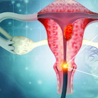
Cervical Cancer
Cancer Conditions
Cervical cancer starts in the cells lining the cervix -- the lower part of the uterus (womb). The cervix connects the body of the uterus (the upper part where a fetus grows) to the vagina (birth canal). Cancer starts when cells in the body begin to grow out of control. To learn more about how cancers start and spread, see What Is Cancer? What Is Cancer? Cancer starts when cells in the body begin to grow out of control. Cells in nearly any part of the body can become cancer cells. Learn more here. Anatomy Gallery: Female Genitourinary System Explore our 3D interactive tour of the female genitourinary system. The cervix is made of two parts and is covered with two different types of cells. The endocervix is the opening of the cervix that leads into the uterus. It is covered with glandular cells. The exocervix (or ectocervix) is the outer part of the cervix that can be seen by the doctor during a speculum exam. It is covered in squamous cells. The place these two cell types meet in the cervix is called the transformation zone . The exact location of the transformation zone changes as you get older and if you give birth. Most cervical cancers begin in the cells in the transformation zone. Cells in the transformation zone do not suddenly change into cancer. Instead, the normal cells of the cervix first gradually develop abnormal changes that can turn into cancer. Doctors use several terms to describe these cell changes, including cervical intraepithelial neoplasia (CIN) , squamous intraepithelial lesion (SIL) , and dysplasia . Y ou might hear these abnormal changes referred to as precancers or precancerous changes. When these abnormal changes in the cervix are found, they are graded on a scale of 1 to 3 based on how much of the cervical tissue looks abnormal. In CIN1 (also called mild dysplasia or low grade SIL ), not much of the tissue looks abnormal. Most often, these cells will change back to normal cells. In CIN2 or CIN3 (also called moderate/severe dysplasia or high-grade SIL ) more of the tissue looks abnormal. With these cell changes, there is a higher risk that the cells can become cancer cells and will need to be watched closely or removed. Although cervical cancers start from cells with abnormal changes, only some women with these cervix changes will develop cancer. For most women, these abnormal cells will go away without any treatment. But, in some women, these abnormal cells can turn into true (invasive) cancers. Treating abnormal changes in cervical cells can prevent almost all cervical cancers. Cervical cancer screening looks for abnormal cells in the cervix or cervical cancer early, when treatment is more likely to be successful. Regular screening can prevent cervical cancer and save lives. The HPV test and the Pap test are used to screen for cervical cancer: The Pap test can detect and treat precancerous changes to prevent cancer from developing. The HPV test looks for infection by high-risk types of HPV that are more likely to cause precancers and cancers of the cervix. There is no treatment for HPV infection, but a vaccine can help prevent it. See Can Cervical Cancer Be Prevented? The specific types of treatment for abnormal screening tests are discussed in When Cervical Screening Test Results are Abnormal . Cervical cancers and cervical precancers are classified by how they look in the lab under a microscope. The main types of cervical cancers are squamous cell carcinoma and adenocarcinoma . Most (up to 9 out of 10) cervical cancers are squamous cell carcinomas . These cancers develop from cells in the exocervix. Squamous cell carcinomas most often begin in the transformation zone (where the exocervix joins the endocervix). Most other cervical cancers are adenocarcinomas . Adenocarcinomas are cancers that develop from glandular cells. Cervical adenocarcinoma develops from the mucus-producing gland cells of the endocervix. Less commonly, cervical cancers have features of both squamous cell carcinomas and adenocarcinomas. These are called adenosquamous carcinomas or mixed carcinomas . Although almost all cervical cancers are either squamous cell carcinomas or adenocarcinomas, other types of cancer also can develop in the cervix. These other types, such as melanoma , sarcoma , and lymphoma , occur more commonly in other parts of the body. Only the more common cervical cancer types are covered here. It is important for you to have frank, open discussions with your cancer care team. They want to answer all your questions to help you make informed treatment and life decisions. Here are some questions to consider. What type of cervical cancer do I have? Has my cancer spread outside the cervix? Can the stage of my cancer be determined and what does that mean? Will I need other tests before we can decide on treatment? Do I need to see any other doctors or health professionals? If I’m concerned about the costs and insurance coverage for my diagnosis and treatment, who can help me? What are my treatment choices? What treatment do you recommend and why? How much experience do you have treating this type of cancer? Should I get a second opinion ? How do I do that? Can you recommend someone? What would the goal of the treatment be? How quickly do we need to decide on treatment? What should I do to be ready for treatment? How long will treatment last? What will it be like? Where will it be done? What risks or side effects are there to the treatments you suggest? Are there things I can do to reduce these side effects? How might treatment affect my daily activities? Will the treatment put me into menopause early? Will I need hormone replacement therapy after treatment? If so, is it safe? What are the chances my cancer will recur (come back) with these treatment plans? What will we do if the treatment doesn’t work or if the cancer recurs? Will I be able to have children after my treatment? What are my treatment options if I want to have children in the future? Once treatment begins, you’ll need to know what to expect and what to look for. Not all these questions may apply to you, but asking the ones that do may be helpful. How will we know if the treatment is working? Is there anything I can do to help manage side effects ? What symptoms or side effects should I tell you about right away? How can I reach you on nights, holidays, or weekends? Do I need to change what I eat during treatment ? Are there any limits on what I can do? Can I have sex during treatment ? Will my sex life change after treatment? What kind of exercise should I do, and how often? Can you suggest a mental health professional I can see if I start to feel overwhelmed, depressed, or distressed ? Will I need a special diet after treatment? Are there any limits on what I can do? What other symptoms should I watch for? What kind of exercise should I do now? What type of follow-up will I need after treatment ? How often will I need to have follow-up exams and imaging tests? Will I need any blood tests? How will we know if the cancer has come back ? What should I watch for? What will my options be if the cancer comes back? Along with these examples, be sure to write down some of your own. For instance, you might want more information about recovery times. You might also ask if you qualify for a clinical trial. Keep in mind that doctors aren’t the only ones who can give you information. Other health care professionals, such as nurses and social workers, can answer some of your questions. To find out more about speaking with your health care team, see The Doctor-Patient Relationship . About Cervical Cancer Key Statistics for Cervical Cancer What's New in Cervical Cancer Research? What Is Cervical Cancer? Causes, Risk Factors, and Prevention Early Detection, Diagnosis, and Staging Treating Cervical Cancer After Treatment Cervical Cancer Quiz Surgery for Cervical Cancer Radiation Therapy for Cervical Cancer Chemotherapy for Cervical Cancer Targeted Drug Therapy for Cervical Cancer Immunotherapy for Cervical Cancer Treatment Options for Cervical Cancer, by Stage Questions to Ask About Cervical Cancer Fertility Seeking a Second Opinion Health Professionals Who Are Part of a Cancer Care Team Source: https://www.cancer.org/cancer/types/cervical-cancer.html